Pharmacophore an International Research Journal
Pharmacological action is conventionally summarized through systemic exposure, tissue-average concentration, target expression, or aggregate response, yet each of these measures can obscure the local conditions that determine whether a drug reaches, engages, and alters a biologically relevant tissue state. This article develops a proposed Spatially Resolved Computational Pharmacology Architecture for representing drug action as an address-dependent process within heterogeneous microenvironments. The approach integrates coordinate-resolved exposure, transport, target accessibility, cell identity, tissue state, target engagement, response, resistance, toxicity, uncertainty, and whole-body coupling while preserving the evidentiary distinctions among measured, inferred, and simulated quantities. The synthesis argues that no single performance metric can establish spatial pharmacological adequacy because local action depends on the interaction of delivery, binding, cellular composition, structural barriers, adaptive dynamics, and temporal context. The central contribution is an architecture that assigns explicit data contracts, uncertainty boundaries, and validation obligations to each component, thereby preventing spatial maps, omics-derived states, and mechanistic simulations from being treated as interchangeable evidence. The article further distinguishes spatial association from mechanism, local exposure from pharmacologically available concentration, target expression from engagement, and benchmark performance from prospective pharmaceutical usefulness. Important limitations include incomplete tissue sampling, cross-platform registration error, uncertain scale translation, sparse longitudinal measurements, parameter non-identifiability, and restricted evidence outside intensively studied disease settings. The proposed architecture is therefore not a validated predictive system or clinical decision tool. Its value lies in organizing testable questions, guiding multimodal evidence integration, and defining the conditions under which localized and systemic therapies may be evaluated with greater spatial and mechanistic discipline.
Introduction
Pharmacological action occurs in organized tissue rather than in an anatomically neutral compartment. Vessels, extracellular matrix, epithelial or tumour structures, immune aggregates, stromal barriers, necrotic regions, metabolic gradients, and organ-specific interfaces determine which molecules reach particular cells and under what physicochemical conditions. Spatial transcriptomics has made tissue architecture an explicit analytical object by locating molecular measurements within their physical surroundings rather than treating them as dissociated averages [1]. This development changes the computational-pharmacology question from whether a drug is present in an organ to where it is present, in what form, near which targets, and within which biological state. The address of an exposure is therefore not an optional annotation added after pharmacokinetic analysis; it can be part of the causal context through which exposure becomes—or fails to become—pharmacological action.
Single-cell measurements have greatly expanded the resolution at which pharmaceutical systems can be described, but cell identity alone does not specify the tissue relationships that govern delivery, signalling, adaptation, or injury. Integrating single-cell and spatial transcriptomics allows molecular states to be interpreted in relation to neighbouring cells, structural compartments, and intercellular dynamics [2]. For computational pharmacology, this distinction is critical. Two cells with similar transcriptional identities may experience different drug concentrations, ligand environments, mechanical constraints, oxygenation states, or immune interactions because they occupy different locations. Conversely, cells that appear molecularly distinct may participate in the same functional niche. An address-resolved representation must therefore encode both the properties of individual cellular units and the higher-order organization through which local pharmacology emerges.
The same problem extends beyond RNA. Pharmacological reasoning often depends on protein abundance, post-translational state, membrane localization, target accessibility, receptor occupancy, metabolic activity, and tissue morphology. Multiplex antibody-based imaging can preserve protein composition and tissue organization within a common spatial context, although its interpretation remains conditional on antibody validation, segmentation quality, panel design, and image-processing choices [3]. Computational and AI-based pharmaceutical models may detect spatial patterns across these measurements, but pattern recognition does not determine whether a feature is a causal transport barrier, a response marker, a correlate of disease severity, or a technical artifact. A high-performing model can therefore reproduce a spatial label while remaining pharmacologically uninformative if its inputs, biological meaning, and context of applicability are not specified.
Spatial multi-omics creates an opportunity to connect molecular layers, cell states, morphology, metabolism, and tissue structure, but it also introduces mismatched resolution, incomplete modality coverage, registration error, batch effects, and uncertainty about whether different measurements refer to the same biological unit [4]. The unresolved problem is consequently architectural as much as analytical: pharmaceutical science lacks a coherent representation that keeps local exposure, transport, target accessibility, engagement, cell and tissue state, response, resistance, and toxicity distinct while connecting them across tissue and whole-body scales. This article proposes a Spatially Resolved Computational Pharmacology Architecture, abbreviated SRCPA, as an original conceptual and methodological contribution. SRCPA is intended to organize coordinate-indexed evidence, modelling relationships, uncertainty, and validation obligations; it is not presented as an empirically validated model, universal ontology, clinical recommendation system, or deployment-ready platform.
Why Location Determines Pharmacological Effect
The first reason location matters is that drug presence is not spatially uniform and cannot always be inferred from plasma exposure or a homogenized tissue sample. Mass spectrometry imaging can reveal regional distributions of administered compounds and metabolites while retaining histological context, thereby exposing gradients, reservoirs, excluded regions, and chemically distinct microdomains that tissue-average measurements may conceal [5]. A local signal, however, is not automatically equivalent to unbound, pharmacologically available concentration. Ionization efficiency, matrix effects, spatial resolution, tissue processing, chemical identification, and sampling time can alter the observed map. SRCPA therefore defines a local exposure field as a coordinate- and time-indexed representation whose provenance must distinguish measured signal, calibrated concentration, inferred free fraction, and model-predicted exposure.
The second reason is that large molecules encounter delivery constraints that emerge from three-dimensional tissue organization. Multiplex three-dimensional mapping has shown that macromolecular distribution can vary with vascular access, distance from vessels, binding, and microenvironmental structure [6]. These relationships imply that nominal target abundance may be pharmacologically irrelevant when a therapeutic cannot reach the target-bearing compartment or is sequestered before reaching it. In the proposed architecture, transport and target accessibility are consequently separate components. The transport component represents perfusion, permeability, diffusion, convection, binding, metabolism, and clearance, whereas the target-accessibility component represents whether a target is physically and molecularly reachable under local conditions. Combining them prematurely would obscure whether treatment failure arises from absent exposure, inaccessible target, insufficient engagement, or downstream biological insensitivity.
The third reason is that pharmacological influence may extend beyond the point of initial target engagement. Spatial and temporal analysis of an antibody–drug conjugate has demonstrated that tumour penetration and bystander activity can interact, with payload effects extending from directly targeted cells into neighbouring regions [7]. This illustrates why a spatial exposure map cannot by itself delimit the field of action. Released payloads, diffusible mediators, intercellular signalling, immune recruitment, metabolic transformation, and tissue damage may propagate beyond the original binding site. SRCPA therefore distinguishes the exposure field from an engagement field and a response-propagation operator. The distinction is particularly important for modalities in which the location of binding, intracellular release, active metabolite formation, and biological effect do not coincide.
The fourth reason is that tissue structures can create conserved delivery barriers that are invisible to analyses based only on systemic concentration and target expression. Single-cell spatial pharmacobiology has identified stromal and extracellular-matrix neighbourhoods associated with restricted therapeutic-antibody delivery and local target engagement in human solid tumours [8]. Such evidence supports the proposition that pharmacological opportunity is defined jointly by exposure, transport, target accessibility, and microenvironmental state rather than by any one measurement. Across small-molecule imaging, three-dimensional macromolecular mapping, and single-cell antibody pharmacobiology, the convergent finding is that nominally exposed tissue can contain substantially different local delivery conditions [5, 6, 8]. SRCPA treats this convergence as evidence for an address-resolved representation, not as proof that one transport mechanism, spatial threshold, or predictive model applies universally. Table 1 organizes the evidence, constructs, and interpretation boundaries needed to develop why location determines pharmacological effect within the article’s central argument.
Table 1. Evidence domains, core questions, scientific requirements, and interpretation boundaries for Why location determines pharmacological effect
|
Construct or evidence domain |
Core question |
Scientific basis |
Role in the article |
Failure or overclaiming risk |
Boundary statement |
|
Local drug exposure |
What drug-related material is present at a specified tissue coordinate and time? |
Spatial chemical imaging, quantitative tissue assays, molecular imaging and pharmacokinetic modelling |
Establishes the address-dependent input to pharmacological action |
Treating image intensity, total concentration or metabolite signal as active free drug |
Local detection does not alone establish pharmacologically available exposure |
|
Three-dimensional transport |
How do vessels, matrix, pressure, permeability, diffusion, convection and binding shape delivery? |
Anatomical imaging, vascular measurements, transport experiments and mechanistic models |
Explains how systemic input becomes a heterogeneous tissue distribution |
Inferring a unique mechanism from a fitted distribution |
A plausible transport model requires independent parameter and perturbational support |
|
Target abundance |
Is the molecular target expressed in the local tissue region? |
Spatial transcriptomics, proteomics, multiplex pathology and validated molecular probes |
Identifies candidate regions of pharmacological relevance |
Equating transcript or total protein abundance with reachable target |
Expression is necessary for some mechanisms but does not establish accessibility or engagement |
|
Target accessibility |
Can the therapeutic physically and molecularly reach the relevant target state? |
Subcellular localization, tissue structure, binding-site availability and accessibility assays |
Separates target presence from actionable target opportunity |
Assuming every detected target is equally available to every modality |
Accessibility is modality-, state-, dose- and time-dependent |
|
Target engagement |
Does the drug interact with its intended target at the specified address? |
Occupancy measurements, labelled agents, proximal biomarkers and competitive-binding evidence |
Connects exposure with proximal pharmacological action |
Inferring engagement from colocalization or target expression alone |
Engagement must be measured or supported by an adequately validated mechanistic inference |
|
Effect propagation |
Does action remain local or spread through payload diffusion, signalling or multicellular responses? |
Bystander-effect studies, metabolite mapping, signalling measurements and longitudinal pathology |
Explains why the location of effect may differ from the location of initial binding |
Treating spatial proximity as proof of propagated causality |
Propagation requires temporal and perturbational evidence |
|
Tissue microenvironment |
Which cellular, structural and physicochemical conditions modify delivery and response? |
Spatial omics, pathology, metabolic imaging and mechanical characterization |
Supplies the context in which exposure becomes response, resistance or toxicity |
Assigning causal status to observational neighbourhoods |
A spatial association may identify a candidate modifier but not establish a mechanism |
|
Whole-body–tissue coupling |
How do systemic disposition and local tissue processes constrain one another? |
Plasma pharmacokinetics, organ distribution, physiologically based models and tissue measurements |
Links local pharmacology to dosing and systemic exposure |
Allowing local predictions to violate mass balance or systemic disposition |
Cross-scale coherence is necessary but not sufficient for biological validity |
|
Uncertainty and provenance |
What was measured, inferred or simulated, and how reliable is each spatial element? |
Analytical validation, registration quality, model calibration and metadata |
Prevents evidence categories from being silently conflated |
Presenting one confidence score as comprehensive certainty |
Uncertainty must remain specific to modality, scale, model and context of use |
Spatial Heterogeneity in Exposure, Targets, Cells, and Tissue States
Spatial heterogeneity is not adequately represented by dividing tissue into arbitrary high- and low-expression regions. It arises from organized relationships among cells, structural compartments, molecular gradients, and physical barriers. Multiplexed ion-beam imaging of triple-negative breast cancer has shown that tumour and immune elements form structured spatial patterns rather than random mixtures [9]. For pharmacological modelling, these patterns matter because local immune regulation, stromal contact, vascular proximity, and tumour organization may alter both delivery and downstream response. SRCPA therefore represents a tissue address as more than a Cartesian coordinate. Each address is associated with an evolving state vector that may include exposure, target abundance, accessibility, cell identity, neighbouring populations, structural features, metabolic conditions, and uncertainty. The state vector is proposed as a modelling construct and does not imply that every component can currently be measured in every specimen.
Spatial pathology further demonstrates that heterogeneity exists at multiple nested scales. Single-cell analysis of breast-cancer tissue has identified diverse tumour and stromal phenotypes distributed across distinct tissue contexts [10]. A pharmacological model that reduces this diversity to a dominant cell type or regional mean risks obscuring rare but consequential populations, including target-negative sanctuaries, drug-metabolizing cells, suppressive immune subsets, or repair-associated stromal states. At the same time, increasing resolution does not automatically improve inference. Cellular segmentation errors, marker-panel limitations, sampling bias, and discordance between morphological and molecular classifications can create false precision. SRCPA therefore requires resolution to be selected according to the pharmaceutical question rather than maximized indiscriminately. A model of antibody penetration may require vascular and extracellular-matrix structure, whereas a model of intracellular target engagement may require subcellular localization and target-state measurements.
Higher-order tissue organization must also be distinguished from isolated cellular properties. Coordinated cellular neighbourhoods at the invasive front of colorectal cancer demonstrate that recurrent combinations of cell types and their relationships can define biologically meaningful tissue structures [11]. Single-cell references can additionally be integrated with spatial transcriptomic measurements to infer where fine-grained tumour and stromal states occur within complex tissue architecture [12]. These approaches support two complementary SRCPA components: an observation model that accounts for mixed or incomplete spatial measurements, and a neighbourhood model that represents relationships among localized cellular states. Neither component should convert probabilistic localization into observed ground truth. Deconvolution outputs, neighbourhood assignments, and spatial domains must retain posterior uncertainty, dependence on reference data, and sensitivity to analytical choices.
Localized multicellular immune hubs provide further evidence that tissue function may arise from organized cellular collectives rather than from the abundance of any one population [13]. Accordingly, SRCPA proposes a hierarchy of spatial units comprising measurement pixels, cells, microdomains, neighbourhoods, structural compartments, tissue regions, and organ-level boundaries. Interactions may occur within or across these units, and the relevant level may change with therapeutic modality and timescale. Evidence from structured tumour–immune arrangements, cellular neighbourhoods, and multicellular hubs supports treating tissue organization as a pharmacologically relevant state variable [9, 11, 13]. It does not establish that every recurrent spatial pattern is mechanistic, stable, or therapeutically actionable. The architecture must therefore preserve distinctions among descriptive organization, predictive association, experimentally supported mechanism, and intervention-ready targetability while allowing each evidentiary level to inform appropriately bounded hypotheses.
Architecture for Spatially Resolved Computational Pharmacology
SRCPA begins with a spatial reference frame that assigns tissue coordinates to measured, inferred, and simulated entities without treating them as evidentially equivalent. Giotto demonstrates how spatial expression data can be represented through tissue domains, cellular neighbourhoods, interaction analyses, and coordinate-linked visualizations [14]. In the proposed architecture, this computational layer is extended from descriptive spatial analysis to pharmacological state representation. Each tissue address may contain an exposure state, transport conditions, target abundance and accessibility, cell identity, neighbourhood composition, engagement status, response state, resistance potential, toxicity state, and provenance record. These elements form a structured address-resolved state vector rather than a single latent embedding or composite score. The separation is necessary because two tissue regions may appear similar in one modality while differing substantially in drug availability, target engagement, or vulnerability to injury.
The architecture next requires an observation and localization layer capable of representing uncertainty in cell placement and tissue composition. Cell2location illustrates how single-cell reference profiles can be used to infer fine-grained cell populations within spatial transcriptomic measurements through a probabilistic framework [15]. SRCPA treats such outputs as distributions over possible tissue states rather than as definitive cell maps. Every localized population or state should therefore retain information about reference-dataset suitability, posterior uncertainty, spot or pixel mixture, platform differences, and sensitivity to model assumptions. The proposed architecture also distinguishes biological absence from measurement non-detection. A cell type may be truly absent, present below assay sensitivity, omitted from the reference atlas, or merged into a broader inferred state. These possibilities have different pharmacological consequences and should not be collapsed into the same null value.
A separate measurement-model layer is required because many spatial observations contain mixtures of cells, molecules, and technical effects. Robust decomposition methods demonstrate that spatial transcriptomic measurements may require explicit modelling of cell-type mixtures and platform-specific variation [16]. Within SRCPA, each modality therefore has a data contract specifying its biological entity, spatial support, temporal support, measurement process, preprocessing lineage, and permissible interpretation. A histological region, imaging pixel, transcriptomic spot, segmented cell, mass-spectrometric ion map, and simulated finite element cannot be fused merely because they occupy approximately corresponding coordinates. Integration is permitted only after registration uncertainty, scale mismatch, and entity correspondence have been represented. This design prevents computational convenience from silently converting heterogeneous observations into falsely homogeneous evidence.
Spatial statistics and neighbourhood analyses can then derive local organization, autocorrelation, co-occurrence, exclusion, and boundary features from registered data, as exemplified by the scalable analytical functions implemented in Squidpy [17]. These derived features feed—but do not replace—the mechanistic layer. Cross-scale simulation may subsequently connect tissue-resolved cellular behaviour to systemic pharmacokinetics and disease dynamics. A spatial agent-based tumour model coupled to a whole-patient quantitative systems pharmacology model provides a precedent for exchanging information between local agents and systemic compartments [18]. SRCPA generalizes this coupling as a proposed interface through which whole-body disposition supplies tissue boundary conditions while local exposure, engagement, response, and injury are aggregated back to organ or patient-level states. Mass balance, temporal consistency, and uncertainty propagation are mandatory constraints, but satisfying them does not establish biological completeness or predictive validity.
Table 2 organizes the evidence, constructs, and interpretation boundaries needed to develop architecture for spatially resolved computational pharmacology within the article’s central argument.
Table 2. Components, relationships, uncertainties, and validation needs within Architecture for spatially resolved computational pharmacology
|
Component or layer |
Inputs |
Core function |
Expected output |
Uncertainty or limitation |
Validation requirement |
|
Spatial reference frame |
Histology, imaging coordinates, anatomical landmarks, section metadata |
Registers observations to tissue addresses |
Coordinate system with correspondence and deformation records |
Misregistration, tissue deformation, serial-section mismatch |
Fiducial assessment, landmark agreement and sensitivity to registration choices |
|
Modality-specific observation layer |
Spatial omics, pathology, chemical imaging, molecular imaging |
Preserves the entity and measurement process represented by each modality |
Quality-controlled modality-specific observations |
Detection limits, assay specificity, batch effects and preprocessing dependence |
Analytical controls, reproducibility and orthogonal confirmation |
|
Cell and tissue localization layer |
Single-cell references, spatial measurements, segmentation outputs |
Estimates cell identities, abundances and tissue states at each address |
Probabilistic cell and state maps |
Reference mismatch, mixed pixels and uncertain state definitions |
Comparison with validated markers, pathology review and alternative localization methods |
|
Transport and exposure layer |
Plasma pharmacokinetics, vessels, perfusion, permeability, diffusion, binding, metabolism |
Connects systemic input to local drug and metabolite distributions |
Time-indexed exposure fields |
Parameter non-identifiability, incomplete free-fraction data and model-form error |
Quantitative tissue measurements, mass balance and transport perturbations |
|
Target-accessibility layer |
Target expression, subcellular location, tissue barriers, binding-site information |
Represents whether the intended target can be reached in a relevant molecular state |
Address-specific accessibility estimates |
Transcript–protein discordance and modality-dependent accessibility |
Occupancy, labelled-agent or proximal-engagement evidence |
|
Engagement layer |
Local exposure, accessible target, affinity and competition |
Represents proximal drug–target interaction |
Measured or inferred engagement field |
Indirect biomarkers and temporal mismatch |
Direct occupancy or validated proximal pharmacodynamic measurements |
|
Cell and tissue-state layer |
Cell identity, neighbourhood, metabolism, inflammatory state, mechanics |
Defines the biological context in which engagement is interpreted |
Address-resolved tissue-state vector |
State instability, missing modalities and descriptive classifications |
Repeated measurements, perturbation studies and external tissue confirmation |
|
Response and adaptation layer |
Engagement, baseline state, signalling and temporal information |
Represents desired response and adaptive state transition |
Local response and adaptation fields |
Confounding, sparse time points and uncertain causal direction |
Longitudinal exposure–engagement–response measurements |
|
Resistance layer |
Clonal states, competition, protective niches, treatment history |
Represents emergence, persistence or expansion of resistant states |
Spatial resistance hypotheses or simulations |
Incomplete sampling and unobserved evolutionary routes |
Serial tissue, lineage evidence and treatment perturbation |
|
Toxicity and repair layer |
Off-target exposure, injury markers, histology, organ function |
Represents local damage, stress, compensation and regeneration |
Time-resolved toxicity and repair fields |
Species translation and ambiguity between reversible stress and injury |
Orthogonal pathology, functional markers and cross-species evaluation |
|
Cross-scale coupling layer |
Tissue outputs, organ compartments, whole-body pharmacokinetics |
Exchanges information between tissue and systemic models |
Coherent local and systemic trajectories |
Scale mismatch and compensating parameters |
Conservation checks and independent local and systemic measurements |
|
Provenance and uncertainty layer |
Metadata, model versions, calibration and sensitivity analyses |
Records what is measured, inferred, simulated or proposed |
Address-specific provenance and uncertainty |
Incomplete propagation and misleading global confidence scores |
Calibration against independent observations and full audit trail |
|
Context-of-use validation layer |
Intended question, model influence and consequence of error |
Defines the evidence burden and permissible decision use |
Conditional credibility statement |
Vague use claims and validation detached from consequence |
Predefined acceptance criteria, stress testing and independent evaluation |
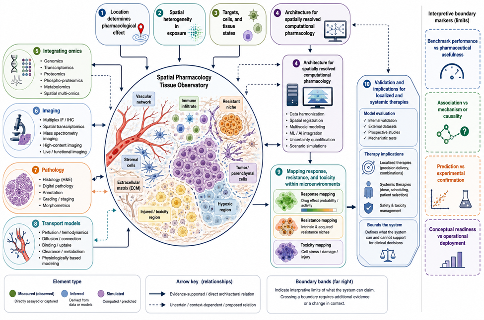
Figure 1 presents the spatial pharmacology tissue observatory, showing how the article’s key components and boundaries are connected within architecture for spatially resolved computational pharmacology.
|
|
|
Figure 1. Spatial Pharmacology Tissue Observatory. |
The figure is an original conceptual synthesis that organizes the article’s central contribution across location determines pharmacological effect, spatial heterogeneity in exposure, targets, cells, and tissue states, spatial heterogeneity in exposure, tissue states, architecture for spatially resolved computational pharmacology, integrating omics, imaging, pathology, and transport models. Arrows and grouping indicate proposed or evidence-supported relationships rather than measured effect sizes. The figure does not represent a validated predictive model, regulatory determination, clinical recommendation, or deployment-ready system.
Integrating Omics, Imaging, Pathology, and Transport Models
Multimodal integration should begin by defining what each modality contributes rather than by maximizing the number of fused features. Deterministic barcoding in tissue demonstrates that RNA and protein measurements can be mapped within a shared spatial coordinate system [19]. For SRCPA, such co-mapping supports joint representation of molecular identity and tissue location, but it does not eliminate modality-specific differences in sensitivity, dynamic range, spatial support, or biological meaning. RNA abundance may indicate transcriptional potential, whereas protein abundance may more closely reflect target availability, although neither alone establishes accessibility or engagement. Integration must therefore retain modality identity and should record whether a relation is directly observed within the same section, transferred across serial sections, inferred from a reference atlas, or simulated.
Metabolic state adds another pharmacologically important dimension because local substrate availability, hypoxia, enzymatic activity, and metabolite accumulation can alter drug activation, degradation, response, and toxicity. The integration of mass spectrometry imaging with mass cytometry demonstrates how metabolite distributions may be coregistered with phenotypically characterized cells within a common tissue section [20]. SRCPA uses this principle to connect chemical and cellular evidence while recognizing that molecular assignment, ion suppression, segmentation, and registration may remain uncertain. A detected metabolite associated with a cell class may reflect production, uptake, extracellular accumulation, or proximity rather than a uniquely identified intracellular pathway. Mechanistic interpretation therefore requires temporal evidence, isotope tracing, perturbation, or orthogonal biochemical measurements where appropriate.
Computational pathology can provide structural and morphological information at scales that are often unavailable from molecular assays alone. Deep learning has been used to relate breast-tumour morphology to spatial gene-expression patterns [21]. Such relationships may help estimate molecular features in unsampled regions or prioritize areas for additional measurement, but predictive concordance does not establish that morphology mechanistically determines expression. Within SRCPA, morphology-derived estimates are therefore labelled as inferred evidence and accompanied by domain-shift and calibration assessments. A model trained on one staining protocol, disease subtype, scanner, or institution may not retain validity under another. Pathology integration should consequently support tissue interpretation and sampling strategy without being presented as a substitute for direct molecular or pharmacological measurement.
Localized therapy illustrates why multimodal integration must also include physical transport. Correlative spectroscopy and mass spectrometry imaging have shown that topical-drug distributions can be interpreted in relation to epidermal and dermal structures [22]. In solid tumours, mathematical and experimental evidence indicates that vascular dysfunction, interstitial pressure, extracellular matrix, and tissue mechanics can constrain penetration and efficacy [23]. SRCPA therefore couples registered molecular and pathological maps to transport models that represent movement rather than simply interpolating between measured pixels. Molecular co-mapping, cell-resolved metabolic imaging, and computational pathology provide complementary views of tissue state, but none can replace the others or independently establish local pharmacological action [19-21]. Their integration becomes scientifically useful only when entity correspondence, spatial scale, temporal alignment, transport constraints, and uncertainty are made explicit.
Mapping Response, Resistance, and Toxicity within Microenvironments
Response should be represented as a spatially and temporally resolved change in biological state rather than as a static label attached to baseline tissue. Spatial multi-omic analysis of glioblastoma has revealed regional tumour–host interdependence involving inflammatory, metabolic, and cellular programs [24]. Such findings support the possibility that identical target engagement may produce different consequences in different microenvironments. SRCPA therefore defines response as a conditional transition from a baseline address-resolved state to a subsequent state under treatment. The transition may include tumour-cell death, pathway suppression, immune recruitment, functional recovery, or another disease-appropriate effect. Observed differences between regions remain associative unless temporal ordering, exposure, engagement, and perturbational evidence support a stronger mechanistic interpretation.
Resistance is likewise spatial because resistant populations compete with sensitive cells and inhabit environments that may either constrain or protect them. Experimental and mathematical work has shown that spatial competition can alter the expansion of resistant cells during targeted therapy [25]. The implication is not that one ecological rule governs every disease, but that the geometry of cell populations and drug exposure can change evolutionary trajectories. In SRCPA, a resistance field represents the probability, abundance, or simulated persistence of resistant states at particular addresses and times. Inputs may include clonal identity, local exposure, neighbouring populations, resource conditions, immune pressure, and prior treatment. Resistance modelling should distinguish pre-existing resistance, induced adaptation, spatial refuge, pharmacokinetic protection, and competitive release because these mechanisms imply different interventions.
Spatial multi-omics has also identified tumour–immune configurations associated with resistance to immunotherapy [26]. These configurations may reveal candidate vulnerabilities, but their pharmaceutical use requires caution. A resistant-region classifier may capture disease severity, biopsy location, treatment history, or technical batch rather than a causal resistance mechanism. SRCPA therefore requires resistance hypotheses to be linked to falsifiable intervention questions: whether altering exposure, removing a barrier, changing a cellular interaction, or targeting a state transition modifies the resistant region as predicted. Where perturbational evidence is absent, outputs should be described as spatial associations or prospective hypotheses rather than mechanistic explanations or treatment-selection rules.
Toxicity must be mapped with the same spatial discipline as efficacy. Mass spectrometry imaging can localize drugs and metabolites in relation to absorption, distribution, metabolism, and toxicological processes [27]. Spatial transcriptomic analysis of cisplatin-associated kidney injury further demonstrates that DNA damage, nephrotoxic responses, and regenerative programs can differ across anatomical regions and time [28]. SRCPA therefore represents toxicity as a dynamic field containing exposure, off-target engagement, cellular stress, tissue injury, compensation, and repair. These states should not be collapsed into one toxicity score because reversible adaptation, molecular stress, structural injury, and organ dysfunction represent different levels of evidence. A spatially favourable efficacy pattern may also coexist with harmful exposure in another organ, requiring benefit–risk interpretation across multiple tissue addresses rather than optimization of the treated lesion alone.
Validation and Implications for Localized and Systemic Therapies
Validation begins with the intended use of the architecture. Imaging, in vitro evidence, and physiologically based pharmacokinetic modelling can provide complementary information for predicting tissue and intracellular drug concentrations [29]. For a localized formulation, the immediate question may concern penetration into a defined anatomical layer; for a systemic small molecule, it may concern target-site concentration relative to plasma exposure; for a biologic, it may concern access to a stromally protected compartment. These questions require different measurements, spatial resolutions, and acceptance criteria. SRCPA therefore rejects a universal validation score. Evaluation must test the specific chain of claims needed for the intended use, including registration, analytical measurement, transport, exposure, accessibility, engagement, response, and uncertainty.
A credible implementation also requires a traceable lifecycle from biological question through model construction and evaluation. Quantitative systems pharmacology workflow principles emphasize explicit questions, biological assumptions, model formulation, calibration, qualification, and iterative refinement [30]. Applied to SRCPA, this means that every component should document why it exists, what evidence informs it, what outputs it may generate, and how failure would be detected. Internal consistency, mass balance, code verification, reproducibility, sensitivity analysis, and parameter plausibility are necessary, but they do not establish that the architecture predicts an unseen tissue or improves a pharmaceutical decision. External and prospective evaluation must be added when the context of use demands stronger evidence.
The burden of validation should increase with the influence of the model and the consequence of an incorrect conclusion. Risk-informed credibility assessment links model evaluation to decision context rather than treating all simulations as equally consequential [31]. An exploratory map used to nominate experiments may tolerate greater uncertainty than a model proposed to exclude a formulation, alter a clinical sampling strategy, or support patient stratification. SRCPA therefore assigns each application a model-influence level, failure consequence, required independent evidence, and human-oversight obligation. This structure does not create regulatory acceptability; it constrains the claims that may reasonably be made from the available evidence.
Mechanistic-model credibility further requires explicit context of use, verification, validation, applicability assessment, and characterization of uncertainty [32]. Independent molecular imaging can provide one form of verification, as illustrated by PET-guided comparison with physiologically based predictions of target-site penetration [33]. For localized therapies, analogous evidence may involve chemical imaging, depth-resolved tissue analysis, or regional pharmacodynamic markers. For systemic therapies, evaluation may require simultaneous plasma, organ, lesion, and intracellular measurements. The central implication is that spatial pharmacology becomes useful not when every address is perfectly observed, but when uncertainty is sufficiently characterized for a bounded decision and competing explanations have been tested. Table 3 organizes the evidence, constructs, and interpretation boundaries needed to develop validation and implications for localized and systemic therapies within the article’s central argument.
Table 3. Evaluation, implementation, and research priorities arising from Drug Action Has an Address Spatially Resolved Computational Pharmacology within Heterogeneous Tissue
|
Evaluation dimension |
What must be tested |
Suitable evidence or assessment |
Failure signal |
Interpretive limitation |
Decision relevance |
|
Analytical validity |
Whether each assay detects and quantifies its intended entity |
Standards, controls, recovery, specificity, replicate concordance and orthogonal assays |
Signal changes with preparation or platform rather than biology |
Analytical validity does not establish pharmacological relevance |
Determines whether a spatial input may enter the architecture |
|
Spatial registration |
Whether modalities refer to the same tissue address |
Fiducials, landmarks, deformation maps and same-section measurements |
Apparent cross-modal relations disappear under plausible realignment |
Low registration error does not establish biological coupling |
Governs whether multimodal integration is permissible |
|
Cell and state localization |
Whether inferred populations and states are correctly placed |
Marker validation, pathology review, alternative models and posterior calibration |
Localization changes substantially with reference or algorithm |
Method agreement may reflect shared assumptions |
Determines confidence in address-specific biological context |
|
Local exposure |
Whether measured or simulated drug distribution is quantitatively defensible |
Calibrated imaging, tissue assays, free-fraction evidence and temporal sampling |
Tissue averages fit while regional predictions fail |
Total abundance may differ from active concentration |
Supports formulation, delivery and target-site questions |
|
Transport mechanism |
Whether distribution is reproduced for the proposed physical reasons |
Mass balance, parameter measurement and barrier perturbation |
Compensating parameters reproduce maps without correct intervention response |
Good fit does not identify a unique mechanism |
Supports delivery-modification hypotheses |
|
Target accessibility and engagement |
Whether the drug reaches and interacts with relevant target states |
Occupancy, labelled agents and validated proximal biomarkers |
Target expression is high but engagement remains absent or unexplained |
Engagement does not establish downstream benefit |
Separates delivery failure from biological insensitivity |
|
Response mapping |
Whether local exposure and engagement precede appropriate biological change |
Longitudinal imaging, pathology, omics and perturbation |
Spatial correlation lacks temporal or dose coherence |
Prediction does not establish causal response |
Supports mechanistic and experimental prioritization |
|
Resistance mapping |
Whether resistant states are detected before or during failure |
Serial tissue, lineage analysis and treatment perturbation |
Model labels only post hoc resistant regions |
Sparse biopsy may not represent whole-tissue evolution |
Supports hypothesis generation for combination or sequencing |
|
Toxicity and repair |
Whether exposure, injury and recovery are correctly distinguished |
Histopathology, toxicogenomics, organ function and temporal assessment |
Stress signatures are misclassified as irreversible injury |
Preclinical tissue patterns may not predict clinical dysfunction |
Supports organ-specific safety investigation |
|
Cross-scale consistency |
Whether local and systemic predictions obey common disposition constraints |
Plasma pharmacokinetics, organ exposure, imaging and conservation checks |
Local trajectories violate dose, clearance or organ balance |
Consistency is necessary but not sufficient for validity |
Connects dosing to tissue-level pharmacology |
|
Uncertainty calibration |
Whether stated uncertainty corresponds to observed error |
External observations, interval coverage, reliability analysis and sensitivity testing |
High confidence accompanies systematic regional error |
Calibration applies only to represented contexts |
Limits the strength of permissible conclusions |
|
Reproducibility and provenance |
Whether results can be regenerated and audited |
Versioned code, metadata, parameter records and independent execution |
Undocumented preprocessing changes the conclusion |
Reproducibility does not prove biological validity |
Enables scientific review and lifecycle governance |
|
Context-of-use credibility |
Whether total evidence is sufficient for the proposed decision |
Risk assessment, predefined criteria, external testing and expert review |
Model influence exceeds supporting evidence |
Credibility is conditional and can change with use |
Determines whether use remains exploratory, supportive or investigational |
Limitations and Boundary Conditions
SRCPA is limited first by the incompleteness of spatial evidence. Most tissue datasets represent a small number of sections, time points, anatomical regions, or patients, while three-dimensional and longitudinal pharmacology may remain largely unobserved. Spatial multi-omics also contains platform-specific gaps, and registration cannot guarantee that serial sections contain identical cells or microstructures [4]. The architecture can represent these uncertainties but cannot recover information that was never measured. Increasing computational complexity may create smoother maps or more detailed latent states without improving biological truth. Biopsy location, tissue handling, disease stage, prior treatment, and assay selection may therefore dominate conclusions unless sampling limitations are explicitly examined.
A second limitation is mechanistic non-identifiability. Similar spatial distributions may arise from different combinations of perfusion, permeability, diffusion, binding, metabolism, cellular uptake, and clearance. Cross-scale coupling can enforce mass balance, yet multiple parameterizations may remain compatible with the same observations, particularly when direct target-site measurements are sparse [18]. SRCPA cannot convert association into causality by architectural declaration. Neighbourhoods, stromal barriers, metabolic states, or morphological features should be treated as candidate determinants until perturbational evidence tests whether changing them alters exposure, engagement, response, resistance, or toxicity. Likewise, a model that predicts spatial patterns accurately may rely on correlates that fail under a new disease, modality, institution, or treatment.
A third boundary concerns decision use. The proposed architecture does not supply dose recommendations, certify target engagement, establish clinical utility, or determine regulatory acceptability. Evidence from single-cell spatial pharmacobiology is compelling for specific solid-tumour and antibody contexts, but it does not prove universal transferability across organs, diseases, therapeutic modalities, or populations [8]. Localized and systemic therapies also pose different validation challenges: ex vivo skin penetration cannot alone establish in vivo benefit, and plasma pharmacokinetics cannot alone validate lesion-level exposure. Human oversight remains necessary to judge biological plausibility, assay fitness, model influence, patient relevance, and consequences of error. SRCPA should therefore be used initially to structure hypotheses, experiments, and evidence rather than to automate high-consequence pharmaceutical decisions.
Research Agenda and Implementation Priorities
The first priority is to establish a common spatial-pharmacology ontology and interoperable data model. Exposure, target accessibility, engagement, tissue state, response, resistance, toxicity, repair, and uncertainty require definitions that remain stable across assays while preserving modality-specific meaning. Benchmark datasets should include matched histology, spatial omics, quantitative drug imaging, pharmacokinetics, and relevant perturbations rather than only cell-type labels or reconstructed expression. Same-section multimodal measurement should be prioritized where feasible, and serial-section studies should report deformation and correspondence uncertainty. Reporting standards should specify coordinate systems, spatial resolution, temporal support, preprocessing, missing modalities, reference atlases, registration procedures, and whether each output is measured, inferred, simulated, or proposed.
The second priority is mechanistic and temporal validation. Studies should pair local exposure with target accessibility, engagement, response, and injury over time so that causal ordering can be evaluated. Barrier-modification experiments, target perturbations, transporter manipulation, local delivery changes, and treatment interruptions can test whether proposed spatial determinants behave as predicted. Resistance studies require serial sampling or imaging capable of distinguishing pre-existing resistant populations from induced adaptation and competitive release. Toxicity studies should integrate molecular stress, histopathology, repair, and organ function rather than treating any isolated signature as definitive injury. External validation should cross disease sites, institutions, preparation methods, therapeutic modalities, and patient populations.
The third priority is staged implementation tied to model risk. Initial use should focus on retrospective tissue interpretation, experimental design, sampling selection, and formulation hypotheses. A second stage may support prospective preclinical studies in which spatial predictions are declared before measurement and tested with independent modalities. Later investigational use may examine whether the architecture improves target-site exposure prediction, combination selection, toxicity investigation, or patient stratification relative to simpler alternatives. Progression between stages should depend on context-specific analytical validity, biological validation, uncertainty calibration, reproducibility, and decision-impact evidence. Infrastructure should support versioned models, provenance-preserving data exchange, auditable workflows, and multidisciplinary review rather than presenting spatial computation as a self-sufficient replacement for pharmacology, pathology, imaging, or clinical judgment.
Conclusion
Drug action has an address because exposure, transport, target accessibility, engagement, cellular state, tissue organization, adaptation, resistance, and injury occur within specific and changing microenvironments. The proposed Spatially Resolved Computational Pharmacology Architecture organizes these determinants as distinct but connected evidence and modelling layers embedded within a common spatial reference frame. Its principal contribution is not a new universal prediction score, but a disciplined structure for preserving what is measured, inferred, simulated, uncertain, and context-dependent while connecting local pharmacology with organ and whole-patient processes. The architecture may support more informative experiments, multimodal analyses, transport models, and validation strategies, but it cannot by itself establish mechanism, clinical utility, dose suitability, regulatory acceptance, or deployment readiness. Its scientific value will depend on quantitative measurement, perturbational testing, longitudinal evidence, uncertainty calibration, cross-scale consistency, and validation matched to the consequence of the intended use.
Acknowledgments: None
Conflict of interest: None
Financial support: None
Ethics statement: None