Pharmacophore an International Research Journal
GASTRIC ULCER HEALING ACTIVITY OF MAHKOTA DEWA “PHALERIA MACROCARPA SCHEFF. BOERL” LEAVES EXTRACT ON MALE WISTAR RATS
Ari Yuniarto*, Ika Kurnia Sukmawati, Vinny Trieagusti
|
|
Pharmacology and Clinical Pharmacy, Bhakti Kencana University, Bandung City, Indonesia |
ABSTRACT
Context: Gastric ulcer is one of the serious problems in human life and has a contribution to mortality in the world. The imbalance between mucosal integrity factors and aggressive factors was known as the pathophysiology of gastric ulcers. Aims: This research aimed to evaluate the gastric ulcer healing activity of mahkota dewa leaves extract (MDLE) on the aspirin-induced rat model. Settings and Design: The animals were divided into 6 groups, each group consisting of five rats. Group I or normal group received the vehicle, group II or control group received aspirin (500 mg/kg body weight (BW). Group III received the standard drug, group IV, V, and VI received 50, 100, and 200 mg/kg BW of MDLE, respectively, and Groups IV to VI received aspirin 500 mg/kg BW and treated using MDLE for three days. After the treatment period, rats were sacrificed and their stomach was incised along the greater curvature. Sucralfate was used as the standard drug for this experiment (90 mg/kg BW). Methods and Material: All reagents in this study were of analytical grade. Aspirin® and sucralfate were purchased from Kimia Farma Pharmacy, Bandung, West Java, Indonesia. The experimental animals were starved for 24h to empty their stomachs and increase the gastric acid level, then a single oral dose of aspirin (500 mg/kg BW) was given to induce gastric injury. 1h before the experiment, water was also withheld. Statistical analysis used: One-way ANOVA, coupled with Dunnet’s test. Results: The results of the study showed that gastric ulcer healing significantly different in the groups received MDLE compared to the control group. Ulcer healing activity of MDLE at the doses of 50, 100, and 200 mg/kg BW was 35.93%, 50.78%, and 66,40%, respectively, and it was observed that 200 mg/kg BW of MDLE was more effective in terms of impact on the number of ulcers, diameter of ulcers, and ulcer index (UI) than other doses. This result was consistent with histopathological analysis, in which 200 mg/kg BW of MDLE could reduce inflammation and improve gastric damage induced by aspirin. Conclusions: It can be concluded that MDLE at a dose of 200 mg/kg BW has potentially antiulcer activity in the aspirin-induced rat model.
Keywords: Aspirin, extract, gastric ulcer, leaves, mahkota dewa.
Introduction
Gastric ulcer is one of the serious problems in the gastrointestinal tract and has a contribution to death worldwide [1]. The most common etiologies of gastric ulcers include Helicobacter pylori infection, long-term use of non-steroidal anti-inflammatory drugs (NSAIDs), and critical illness (stress factor) [2-6].
An imbalance between aggressive and mucosal integrity has been known to be responsible for gastric ulcers. Increased aggressive factors (gastric acid and pepsin secretion) and decreased mucosal integrity factors (prostaglandin, bicarbonate, cellular repair mechanisms, and blood flow in gastric) may lead to gastric ulcers [7, 8]. Gastrointestinal damage associated with the use of aspirin and NSAIDs against the upper gastrointestinal has been reported [9]. The long-term use of aspirin and NSAID may reduce gastric mucosal integrity. Aspirin has two mechanisms to influence gastric damage. The first mechanism by direct irritation in gastric mucosa, and the second mechanism by inhibition of cyclooxygenase (COX) enzyme [10].
Long-term administration of anti-ulcer drugs has been reported to be associated with several side effects. So, the present study aimed to evaluate a natural product, which has potential anti-ulcer activity, used especially in Indonesia. According to several investigations, the use of natural product sources in the experimental animals has shown promising results in the treatment of gastric ulcers [11-14].
Mahkota dewa (Phaleria macrocarpa Scheff. Boerl) is a medicinal plant with various pharmacological activities. It can be found in Indonesia and Malaysia. Its extract has been reported to have pharmacological activities such as anti-inflammatory, anti-diabetic, anti-cancer, anti-bacterial, and antioxidant [15-18]. Therefore, the present study aimed to evaluate gastric ulcer healing activity of mahkota dewa leave extract in the aspirin-induced rat model.
Subjects and Methods:
Experimental Material
All reagents were of analytical grade. Aspirin® and sucralfate were purchased from Kimia Farma Pharmacy, Bandung, West Java, Indonesia.
Plant Material and Identification
Mahkota dewa leaves were collected from Bandung, West Java, Indonesia, and confirmed at Herbarium of Jatinangor, Laboratory of Plant Taxonomy, Department of Biology, Padjajaran University, Indonesia.
Preparation of Extract
Mahkota dewa leaves were dried and ground into powder, then, extracted by the maceration method, evaporated by Rotary Evaporator, and stored for further studies.
Phytochemical Screening of the Extract
Phytochemical screening of MDLE was performed to evaluate the presence of flavonoids, alkaloids, saponins, tannins, quinones, and steroids/triterpenoids.
Experimental Animal
Healthy adult male Wistar rats (200-250g) were used for this experiment. Rats were maintained in a controlled room (standard temperature and humidity) with free access to water and food. The experimental study was performed after approval by the Health Research Ethics Committee, Faculty of Medicine, Padjajaran University.
Induction of Gastric Ulcers
The experimental animals were starved for 24h to empty their stomachs and increase the gastric acid level, then a single oral dose of aspirin (500 mg/kg BW) was given to induce gastric injury. 1h before the experiment, water was also withheld.
Experimental Design
The animals were divided into 6 groups, each group consisting of five rats. Group I or normal group received the vehicle, group II or control group received aspirin (500 mg/kg body weight (BW). Group III received the standard drug, group IV, V, and VI received 50, 100, and 200 mg/kg BW of MDLE, respectively, and Groups IV to VI received aspirin 500 mg/kg BW and treated using MDLE for three days. After the treatment period, rats were sacrificed and their stomach was incised along the greater curvature. Sucralfate was used as the standard drug (90 mg/kg BW).
The gastric juice was collected, centrifuged at 2000rpm for 10 min. The acidity was measured through a digital pH meter. Furthermore, the severity of ulcers was observed and scored to determine the ulcer index. Severity score was given according to Gupta et al [19] (0=normal, 0.5=redness, 1=spot ulcer, 2=hemorrhagic ulcer, 3=deep ulcer, 4=perforation). Ulcer index was determined using the following formula of Vogel [20].
UI = UN +US +UP x10-1
UN = Average of number of ulcers per animal
US = Average of severity score
UP = Percentage of animals with ulcers
The percentage of ulcer healing was determined as: Ulcer healing (%) = (UI control-UI test group)/UI control group × 100%.
Histopathological Study
At the end of the study, the animals were sacrificed and their stomachs were collected, washed with normal saline solution, and kept in 10% formalin buffer solution. Then they were processed using the paraffin method, and stained with hematoxylin-eosin.
Statistical analysis
Statistical analysis was performed using the ANOVA method, coupled with the post-hoc Dunnet’s test. A value of p<0.05 was used to denote statistical significance. All data were expressed as mean ± standard deviation (SD) for each group.
Results and Discussion:
Phytochemical Screening of Extract
The phytochemical screening of MDLE revealed the presence of alkaloids, flavonoids, saponins, tannins, quinones, and steroids/triterpenoids, which may be responsible for gastric ulcer healing.
Aspirin-induced Ulcers
Gastric ulcer is a destruction in the gastrointestinal mucosa that extends to deeper layers through the muscularis mucosa. Helicobacter pylori or non-steroidal anti-inflammatory drugs (NSAIDs) are contributed to gastric ulcers [21]. NSAIDs (e.g. aspirin) are among the most frequently-used medicines [22]. However, their long-term administration may be associated with mucosal injury in the stomach and duodenum. In this study, gastric ulcers were induced by aspirin. As shown in Table 1, all test groups exhibited ulcer healing greater than the control group. Ulcer healing activity of MDLE at the doses of 50, 100, and 200 mg/kg BW was 35.93%, 50.78%, and 66.40%, respectively, and it was observed that 200 mg/kg BW of MDLE was more effective in terms of impact on the number of ulcers, diameter of ulcers, and ulcer index (UI) (2.00 ± 0.70; 1.5±0.40; 5.37±0.80, respectively) than other doses. This result was consistent with histopathological analysis, in which 200 mg/kg BW of MDLE could reduce inflammation and improve gastric damage induced by aspirin. Furthermore, gastric acid pH was another important aspect of this study. As shown in Table 1, MDLE at a dose of 200 mg/kg BW had a significant effect on increasing pH.
Table 1. Effect of MDLE on ulcer healing.
|
Gastric pH |
Number of ulcers |
Diameter of ulcers (mm) |
UI groups |
Ulcer healing (%) |
|
|
Normal group |
3.43±0.39 |
0 |
0 |
0 |
- |
|
Control group |
2.93±0.50 |
4.25±0.5 |
2.25±0.50 |
16.00±0.57 |
0 |
|
Sucralfate 90 mg/kg BW |
3.98±0.60 |
1.75±0.28* |
1.25±0.28* |
3.00±0.57 |
81.25 |
|
MDLE 50 mg/kg BW |
3.12±0.35 |
2.75±0.5* |
1.87±0.25* |
10.25±1.01 |
35.93 |
|
MDLE 100 mg/kg BW |
4.17±0.90 |
2.37±0.75* |
1.75±0.28* |
7.87±1.08 |
50.78 |
|
MDLE 200 mg/kg BW |
4.91±0.50 |
2±0.70* |
1.5±0.40* |
5.37±0.80 |
66.40 |
(*) Significantly different compared to the control group (p <0.05).
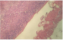
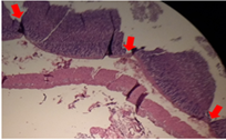
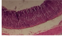
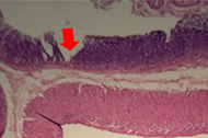
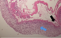
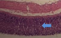
The results of this study showed that MDLE at the dose of 200 mg/kg BW significantly reduced inflammation and influenced stomach tissue regeneration compared to the control group. MDLE at the dose of 50 and 100 mg/kg BW were insufficient to reduce inflammation and tissue regeneration (red arrow: perforation of stomach tissue; blue arrow: tissue regeneration; black arrow: inflammation). Furthermore, sucralfate-treated rats did not show severe destruction in the stomach.
 |
 |
 |
|
Normal group |
Control group |
Sucralfate 90 mg/kg BW |
 |
 |
 |
|
MDLE 50 mg/kg BW |
MDLE 100 mg/kg BW |
MDLE 200 mg/kg BW |
Fig.1 Effect of mahkota dewa leaves extract against stomach histopathological.
Conclusion
Mahkota dewa (Phaleria macrocarpa Scheff. Boerl) is a medicinal plant with various pharmacological activities. Extracts of this plant have been used in traditional medicine to treat several diseases. The experimental study showed that mahkota dewa leaves extract (MDLE) with 200 mg/kg b.w. dose has potentially antiulcer activity on aspirin-induced rats.
Acknowledgment:
This research was funded by Internal Research Grant from LPPM Bhakti Kencana University, Bandung, West Java, Indonesia.
References