Pharmacophore an International Research Journal
This review focuses on the use of state-of-the-art nanotechnology in restorative materials, presenting many innovative approaches. With an eye on improving dental caries management, our findings point to a bright future for creating novel, superior biomaterials. Dental materials have benefited from the novel use of nanotechnology, which offers the possibility of creating materials with superior qualities and antibacterial abilities. In this overview, we review the development of functional nanoparticles and their potential uses in dental restorative materials as helpful tactics for managing dental caries. Additionally, we provide an outline of the suggested remineralizing and antibacterial processes. A systematic literature review from 2000 to 2023 was performed using PubMed, Medline, and ScienceDirect databases. The keywords used were "nanotechnology," "dental caries," and "restorative material." Nanomaterials have considerable promise in reducing the formation of biofilms, blocking the demineralization procedure, remineralizing tooth structure, and fighting microorganisms linked to dental cavities. These promising findings pave the way for further clinical research to determine the restoration products constructed using nanotechnology and their therapeutic efficacy.
Introduction
Dental caries is still the most prevalent and extensive biofilm-dependent oral illness. Cariogenic bacteria, including Streptococcus mutans, Streptococcus mutans, and Lactobacillus, attack teeth, destroying tooth structure in the process. These microorganisms are found in collections of cells attached to the teeth' surface (also known as dental plaque or oral biofilm) [1, 2]. Consequently, caries is a disease that is site-specific and dynamic and arises from an imbalance in the physiologic equilibrium between the mineral ions found in tooth structure and the fluid found in dental plaque, as reflected by the processes of demineralization and remineralization [1]. Tooth demineralization may result from the loss of calcium and phosphate (from enamel and dentin) in the oral biofilm owing to bacterial acids producing pH dips (<5.5). As soon as the toothbrush removes the biofilm, salivary calcium and phosphate may directly work on the tooth to replenish the calcium and phosphate the enamel has lost [3]. There is a little loss of minerals overall since the number of ions obtained is less than the amount lost. Mineral breakdown events in enamel will occur repeatedly if biofilm formation and acid generation are not prevented (demineralization). The disease's clinical symptoms, known as caries or carious lesions, will finally manifest when this surpasses the ability of oral fluids to restore mineral loss [4]. These lesions vary from cavities indentin to white spot lesions, which are early caries lesions that resemble white, chalky patches on enamel. Because this is the causal component, mechanical or nonspecific management of the dental plaque has historically been the focus of efforts to prevent dental caries illnesses.
Finding composite materials that can replicate missing dentinal tissue and restore the original dental appearance is a problem that most dentists encounter. A more recent method of producing sophisticated nanomaterials is creating a bio-mimetic technique based on nanotechnology to mimic natural biomaterials. The way oral antibacterial materials function is by either dissolving or inhibiting the growth of biofilms on the surfaces of the teeth. Incorporating components like titanium, gold, or silver nanoparticles into the biomaterials mixer may enhance their antibacterial characteristics. The broad surface area that metallic nanoparticles (also known as metallic NPs) provide increases their antibacterial potential.
Furthermore, metallic nanoparticles improve mechanical attributes, including toughness and longevity. Dental nanocomposites may benefit from using inorganic nanoparticles, which are mainly based on metal and metal oxide nanoparticles. Pathogenic microorganisms like Streptococcus mutans (S. mutans) may establish colonies and eventually cause tooth decay in the spaces between dental restorations and enamel. Such antimicrobial elements may significantly increase the effectiveness of dental restorative materials [5].
It is possible to regulate the development of cariogenic oral biofilms by using functional materials or structures at the nanoscale (0.1–100.0 nm): nanoparticles may carry bioactive chemicals and antibiotics [6]. Tooth plaque affects the transmission of particles through bio-films by having channels and a vacuum that may go all the way through the biomass of the biofilm to the tooth surface below. In this context, the nanoparticles exhibit potential use due to their adjustable surface charge, degree of hydrophobicity, surface area to biofilm mass ratio, and capacity to adsorb or aggregate on the biofilm surface [6].
Nanotechnology has been instrumental in mitigating the adverse effects on curing reaction kinetics, mechanical strength properties, conversion, unsatisfactory biocompatibility, poor functional performance and aesthetics, and challenging workability. These include challenges related to the conventional size incorporation of antibacterial and remineralizing agents in direct restorative materials. Nanomaterials exhibit superior antimicrobial activity and comparable physical properties compared to traditional materials. This is likely due to the nanoparticles' small size and high surface area, which allow them to release high ions at low filler levels and allow for the incorporation of non-releasing but reinforcing fillers in the same material [6].
Materials and Methods
A systematic literature review from 2000 to 2023 was performed using PubMed, Medline, and ScienceDirect databases. The keywords used were "nanotechnology," "dental caries," and "restorative material."
Inclusion Criteria
Exclusion Criteria
Results and Discussion
Mechanisms of Caries Progression and Prevention
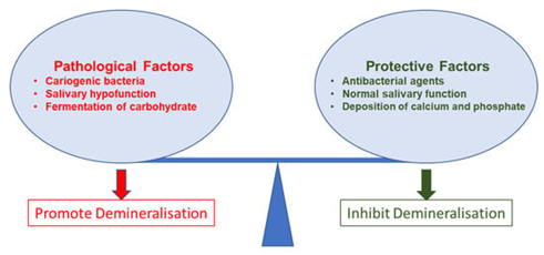
Dental caries has well-established processes. Research has often detailed the demineralization of tooth hard tissue and the impacts of microorganisms (biofilm and cariogenic bacteria) in relation to the caries process. However, these pathways are not linear since caries progression is a complicated process. Cariogenic bacteria, often known as dental plaque, proliferate on surfaces in organized groups. In essence, this tooth plaque is a biofilm. Biofilm causes the development of caries. Precepted biofilm on a tooth's surface does not indicate that caries are present. Only after a complicated interplay of host variables, such as the surface of the tooth, which serves as a stagnation region, fermentable carbohydrates, or free sugars, and cariogenic bacteria that may eventually cause caries expression, does caries expression begin, as shown in Figure 1 [1].
|
|
|
Figure 1. Factors, both preventive and pathological, that influence the demineralization. |
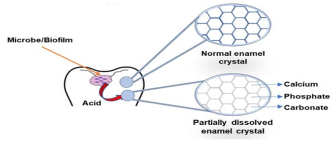
The tooth's crystalline mineral structure demineralizes due to organic acids generated by biofilm bacteria. The primary acid thought to be involved in caries production is lactic acid, which predominates in this process [1]. The pH level falls to a favorable level for the disintegration of hydroxyapatite in tooth hard tissue under these acidic conditions. In an acidic environment, hydrogen ions dissolve hydroxyapatite to produce water, phosphate ions, and calcium ions. Consequently, the tooth's surface demineralizes [7]. Subsequently, the loss of minerals causes enamel crystal derangement, the development of permeability and porosity, and further acid absorption into enamel pores. Acid diffusion further dissolves the hydroxyapatite and lowers the pH near the enamel crystals. Several antibacterial treatments restrict the development of cariogenic bacteria to reduce the amount of organic acids that bacteria create. When salivary calcium and phosphate concentration rises, several antiseptic agents help to restore mineralization by shielding the surface layer from further demineralization [8].
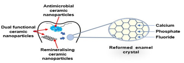
Saliva buffering is essential for maintaining an alkaline pH in the oral environment (Figure 2). Increased pH causes phosphate and calcium ions to re-deposit, which stops demineralization and replenishes the dissolved enamel surface with minerals. As a result, the surface and partly disintegrated enamel crystal remineralize. For teeth to remain intact and undergo remineralization, saliva is necessary. Supplementing saliva with antibacterial/antibiofilm or remineralizing mineral components, or their combination, may stop the development and advancement of dental caries. It can halt demineralization and promote remineralization. Figure 2 shows a schematic image that describes how de- and remineralization work [9].
|
|
|
a) Demineralisation |
|
|
|
b) Remineralisation |
|
Figure 2. Diagram of enamel demineralization and remineralization. (a) Demineralization: Biofilm acid dissolves enamel and releases carbonate, phosphate, and calcium. Antibacterial, remineralizing, or dual-functional ceramic nanoparticles inhibit cariogenic biofilm and restore minerals by collecting calcium, phosphate, and fluoride in partially fragmented enamel crystals. Calcium based Nanoparticles |
Calcium comes from dietary sources and is necessary for bones and teeth. In contrast to acidic desolation, the calcium leak also initiates the tooth's demineralization process, which leads to dental caries. Saliva with added calcium helps the body remineralize. As a natural defense against tooth cavities, calcium and phosphate ions primarily inhibit demineralization and enhance remineralization. Nonetheless, the equilibrium of these ions disturbs and reorganizes the enamel surface in the chronic cariogenic state [1]. Another source may satisfy the need. Remineralization, a noninvasive method of managing dental cavities, has shown to be very beneficial in clinical dentistry. To meet these needs, several researchers have used calcium nanoparticles. To manage dental caries, researchers have recently examined several calcium phosphate-based remineralization systems [6].
The architecture and chemical makeup of hydroxyapatite, a naturally occurring calcium phosphate apatite, are comparable to those of human hard tissues. The primary substance that makes up teeth and bones is hydroxyapatite. Its typical dimensions are 60 nm in length and 5–20 nm in breadth. It gives hard dental tissue's fundamental structure its strength and stiffness. Since nanohydroxyapatite resembles bone and teeth in both morphology and mineral structure, it has drawn much interest and shows promise in cardiology research. Owing to its antibacterial action, biocompatibility, and bioactivity, it may augment some advantageous characteristics of current restorative materials [10].
Research incorporating nanohydroxyapatite into toothpaste or restorative formulations has shown the material's potential for remineralization. Nanohydroxyapatite was efficient in remineralizing caries lesions. Under dynamic pH-cycling circumstances, research examining various doses of nanohydroxyapatite on early enamel caries lesions discovered that nanohydroxyapatite enhanced surface microhardness. Research also claimed to show the formation of new layers on the surface of demineralized enamel due to the deposition of nanohydroxyapatite particles on its cellular structure [11].
Glass-Based Bioactive Nanoparticles
A common bioceramic material in biomedical applications is bioactive glasses, which are surface-reactive materials. They often break down in bodily fluids to generate apatite crystals that demonstrate chemical connections to the surface of the tooth and bone and mimic tooth and bone tissue. Research using nano bioactive glass, including resin composites, revealed a consistent apatite layer growth on the tooth surface without impairing the underlying characteristics of the materials. Meanwhile, a composite incorporating nanobioactive glass raised demineralized dentin's microhardness. Nanobioactive glass may promote the development of hydroxyapatite and osteoinductive properties.
Regarding oral diseases, nanobioactive glass lowers dentin permeability and sensitivity, plugs the orifices of the dentinal tubules, and generates hydroxyapatite on the dentin surface [12]. The demineralized dentin that underwent treatment with nano bioactive glass exhibited an enrichment of minerals and ions. The surface of the carious lesion additionally became more microhardic due to the nanobioactive glass [13].
Despite a lot of coverage, researchers still need to dig deep into remineralization. Our data does not explicitly indicate whether this mineralization is intrafibrillar or extrafibrillar. One area of research was the conjugation of aspartate, serine, glycine, and arginine using nanobioactive glass. The demineralized dentin matrix exhibited the formation of a crystal lattice, as demonstrated by the findings. Intrafibrillar mineralization and cohesive strength are greatest in dentin with a crystal lattice structure. For these reasons, scientists have suggested this composite for use in bonding interfaces, regenerative dentistry, hypersensitivity, and the treatment of dentin erosion [4].
Nanoparticles Based on Silica
Silicon dioxide makes up the inorganic ceramic substance silica. Amorphous fine silica particles disperse insolently to form silica in a colloidal solution. One of the appealing minerals for collagen penetration is silica. Presumably, it can pass through the demineralized collagen matrix without causing surface precipitation. Mesoporous silica nanoparticles doped with calcium, used as inorganic fillers, enhance the mechanical characteristics of resin composites. According to some research, these nanoparticles can be used as a carrier for ciprofloxacin hydrochloride to give antibacterial properties and help prevent secondary caries [3, 6].
Consequently, the scientists hypothesized that these nanoparticles had the potential to lessen tooth erosion. In previous investigations, researchers have looked at the effects of silica nanoparticles and nanohydroxyapatite on erosive enamel and dentin lesions. They stated that there was no statistically significant difference in the mineral deposition in enamel. On the other hand, compared to nano silica infiltrant, nanohydroxyapatite considerably increased mineral infiltration in dentin [2, 14].
Nanotechnology-Based Techniques for Managing Dental Caries
Antimicrobial Strategy
The exceptional antibacterial capabilities of metal nanoparticles (such as zinc and silver) and antimicrobial polymers have attracted a lot of attention over the years [15]. Because of their high surface area-to-volume ratio, which enables a more substantial atom concentration on the surface and maximum environmental contact, these nanostructured agents exhibit strong antibacterial characteristics. Furthermore, these particles' tiny size facilitates better penetration across cell membranes, influencing intracellular processes and raising reactivity and antibacterial activity [12]. This is especially useful since, in comparison to planktonic diseases, microorganisms in biofilms are more resistant to antibacterial agents, and therapy may need considerably more concentrated biocides [16]. Because of this, nanotechnology has allowed for a renewed investigation and improvement of the biologic properties of antibacterial drugs with broad-spectrum actions, safety problems related to dosage, and toxicity toward host cells. The next section reviewed relevant antimicrobial chemicals that have been documented in the literature, their proposed modes of action, and their current use in direct restorative materials.
Nanoparticles of Silver
Numerous antimicrobial applications, including implant coatings and wound dressings, have made use of silver nanoparticles. [16], but their specific mechanism of action is still a mystery. For antimicrobial purposes, silver's efficacy depends on two factors: the amount of bioactive (Ag+1) generated and their interaction with bacterial cell membranes [17]. Smaller silver nanoparticles have a more substantial bactericidal effect than bigger silver nanoparticles because their increased surface area allows more atoms to interact with their surroundings [18, 19]. Silver ions kill bacteria by interacting with their plasma membrane and peptidoglycan cell wall. Silver ions interact with exposed SH groups of proteins, especially those of enzymes involved in vital cellular processes, such as the electron transport chain, inhibiting bacterial DNA replication [19].
Table 1. Methods for preventing dental caries and oral biofilms using nanotechnology
|
Nanotechnology-based agent |
Action |
Benefiting restorative material |
Refs |
|
NAg |
Antimicrobial |
Composite resin; |
[18] |
|
NZn |
Antimicrobial |
Composite resin |
[19] |
|
QAS-PEI |
Antimicrobial |
Composite resin; |
[6] |
|
(CaPO4) nanoparticles |
Remineralizing |
Composite resin; |
[20] |
|
(CaF2) nanoparticles |
Remineralizing |
Composite resin |
[21] |
|
nano-HA, nano-FHA |
Remineralizing |
Resin-modified glass ionomer cement |
[5] |
Zinc Oxide Nanoparticles
Similar to silver, ZnO has shown antibacterial characteristics against several bacteria, including S. mutans. Research has shown that NZn is more effective than traditional particles in killing both Gram-positive and -negative bacteria. In order to inhibit the growth of plankton bacteria, NZn causes changes in cell membrane function and oxidative damages, which in turn generate active oxygen species such as hydrogen peroxide. In gram-negative microbes, ZnO increased transcription levels of genes associated with oxidizing and general stress by a ratio of 3 to 52. There is little effect on human cells from this NZn, but they mainly affect microorganisms (Table 1).
Another potential antibacterial action of NZn is the leaching of zinc ions into the growth medium. This hinders sugar active transport and metabolism and disturbs the functioning of enzymes by displacing Mg2+, which is essential for the metabolic processes of dental biofilms. On S. mutans strains, NZn exhibited antibacterial properties; however, its effectiveness needed a greater concentration than that of NAg. 10% NZn content in dental composites moderately lowers bacterial populations and biofilm formation. On the other hand, the antibacterial effectiveness was not as high as it was for the composite containing NAg. Recent research looked at the characteristics of various NAg (0–5%) resin composites. The growth of S. mutans was examined using the direct contact test. The results showed that the weight (%) of ZnO significantly reduced the development of the bacteria; however, after 24 hours, no discernible antibacterial effect was found. Most of the examined mechanical and physical characteristics were unaffected by adding NZn. In order to demonstrate antibacterial activity in dental composites without compromising mechanical qualities, future studies should focus on appropriate rates of nanofiller fraction [22].
Nanoparticles of Quaternary Ammonium Polyethyleneimine
Dental materials now comprise polymers that include quaternary ammonium (QAS) salts [17]. The benefit of QAS is that the antibacterial agent is immobilized in the composite and not released or lost over time since it copolymerizes with the resin by building a covalent link with the polymer network. This process gives teeth a long-lasting and permanent antibacterial property without appreciably altering the oral cavity's biological balance. After six months of water aging, adhesive systems using QAS showed comparable antibiofilm capabilities. On the other hand, dental composites made by just mixing organic fluoride salts with dental monomers tend to create ion pairs that might leak out. Over time, leaching causes increased water sorption and solubility and diminished mechanical characteristics, shortening these materials' clinical lifespan [21].
Using Biomimetic Techniques
The term "biomimetic products" refers to manufactured materials, structures, or techniques that emulate or mimic biological systems. Combining the ideas of nanotechnology with biomimetic tactics may provide new perspectives and innovative methods for treating and preventing dental caries. Mixing mineral nanomaterials or ion solutions with various organic elements, chemicals, or surfactants makes it feasible to reproduce the ordered nanostructures of tooth enamel [13].
The present research uses an innovative method of treatment: the production of biomimetic dental enamel by using Ca3 (PO4)2 nanoparticles. Chelation of Ca2+ by amelogenin and CPP-ACP is the primary mechanism by which dental hard tissues undergo biomineralization. Investigations have also been made into chitosan's cariogenic and chelating properties [21]. CPP-ACP mimics salivary function by retaining more phosphate and calcium than milk [20]. Research on CS-AMEL hydrogel for preventing tooth decay and restoring erosive lesions is underway. The amelogenin-chitosan hydrogel-repaired enamel exhibited much greater stiffness and elastic modulus than the control samples. This was due to the presence of structured bundling of apatite tiny crystals, which mimicked the structure of enamel [23, 24].
Table 2 briefly summarizes the biomimetic methods that use nanomaterials for enamel manufacturing. In addition,
Table 2. Research on dental cavities using nanoparticles has shown exciting results.
|
Combined strategy |
Key finding |
Ref. |
|
F-HAP |
Repairing early caries lesions and reinforcing the natural enamel are two goals of the nanocrystalline growth technique that F-HAP material may accomplish without excising the tooth. |
[21] |
|
Rod-shape apatite nanocrystals |
It is now known that fluoride hydroxyapatite nanocrystals may remineralize the enamel surface by creating a mineral layer. Fluoride and amelogenin played a supporting role in controlling the size, orientation, phase, and habit of the calcium-phosphate crystals, forming tiny rod-shaped apatites that looked and felt much like natural dental enamel. |
[22] |
|
Pchi–ACP |
Comparable to fluoride, Pchi-ACP remineralized enamel lesions. The rate of remineralization was much more significant with Pchi-ACP therapy compared to fluoride treatment. |
[25] |
|
CS-AMEL |
Since the CS-AMEL hydrogel contains structured bundles of apatite crystals that mimic the structure of enamel, the restored enamel outperformed the control samples in terms of stiffness and elastic modulus. |
[23] |
|
CPP-ACP |
CPP-ACP is comparable to statherin in many ways and can maintain calcium and phosphate concentrations that are much greater than on milk. This makes them a potential salivary biomimetic. |
[23] |
|
Polymer coated liposomes |
Liposomes coated with pectin did not form aggregates when exposed to saliva. Because of this, they show promise as oral medication delivery methods. Dental enamel and HA were both adsorbed by pectin-coated liposomes. Both charged liposomes covered with pectin, and uncoated liposomes could adhere to enamel surfaces at modest shear strengths. |
[26] |
|
Hydroxyapatite nanorods with surfactants |
To create highly ordered enamel prism-like superstructures floating on water, synthesize hydroxyapatite nanorods and modify their surfaces using surfactant monolayers. This will give the nanorods unique surface features. The size and chemical makeup of the artificial hydroxyapatite nanorods are similar to those of natural enamel crystals. |
[26] |
The Limitations in the Use of Nanoparticles
Despite compelling findings from research on the use of nanomaterials in dentistry, there is still much worry about its biosecurity. Furthermore, these worries have changed due to the growing usage of nano biomaterials, with the high absorption rate being seen as the primary issue. A large surface area to volume ratio has made nanoparticles distinctive, enabling improved absorption via the skin, gastrointestinal system, and lungs. When non-biodegradable nanoparticles build up in various human organs, biological tissues may experience adverse side effects. Moreover, current research has shown that nanoparticles may reside in the central nervous system and penetrate the blood-brain barrier. Therefore, understanding the possible neurotoxic effects of various nanomaterials is a crucial first step in using nanotechnology for medicinal applications.
Apatite Nano Hydroxy and Nano Fluoro Hydroxy
One viable alternative to dentin's naturally occurring mineral component is synthesized hydroxyapatite (HA), which is biocompatible and an acceptable substitute. Researchers have looked at using HA powders in biomaterials for the recovery of minerals effects and mechanical property improvement in restorative dental components due to their remarkable biological compatibility and biological activity. To make materials that mend like human hard tissues, researchers combined RMGIC with nano-HA particles. 10% nano-hydroxyapatite (60-100 nm) applied to glass ionomer cement enhanced the bonding ability and resistance to mineral removal compared to micro-HA. The glass ionomer cement incorporating nano-HA, however, took longer to set than the maximum setting duration that is clinically appropriate. The compressive, diametral tensile, and biaxial flexural strengths of glass ionomer cement, which includes nano-hydroxyapatite and nano-FHA, were higher than those of the control.
The possibility of a greater fluoride release through glass-ionomer cement is further enhanced by the fluoride present in nano-fluorohydroxy apatite. Bioactive glass nanoparticles have been suggested for use in restorative materials as a filler and for remineralizing human dentin in a number of investigations. Nanotechnology will play a pivotal role in future treatments due to the higher recovery of minerals rates observed compared to tiny activated glass molecules [27, 28].
Visit Toxicology and Potential Biological Effects
The goal of nanoparticles is to destroy bacteria specifically while avoiding harming human cells. Research has shown a favorable relationship between cytotoxic effects and Silver nanoparticle levels. However, when silver nanoparticles were added to polymers at concentrations of 0.05–0.70%, they were shown to have no cytotoxic effects on human cells. Experimental studies have used doses of Silver nanoparticles ranging from 0.08 to 0.10% mass fraction [23, 29, 30]. They are deficient concentrations compared to a 10% concentration that may cause cytotoxicity in human cells. Limitations: Silver nanoparticles may influence the degree of monomer conversion in dental materials and enhance the quantity of functional residual monomers from the hardened composite [27]. Specific monomers that elute from composites have the potential to produce reactive oxygen species via metabolization or allergic responses [31, 32].
Because it is miscible with the usual DMA subunits used in composite structures, the low-viscosity of the substance quaternary ammonium sulfanilamide has less of a propensity to leach subunits. Comparing the bonding agent with the non-antibacterial commercial version, human cells were not significantly cytotoxically affected by adding 0.5% silver nanoparticles or 10% quaternary ammonium methacrylate. Clarifying the dangers presented by the nanoparticles used in dental materials is essential to advancing their usage, as research on the toxicity of nanomaterials is still in its nascent stages [29, 33].
Conclusion
This review focuses on the use of state-of-the-art nanotechnology in restorative materials, presenting many innovative approaches. With an eye on improving dental caries management, our findings point to a bright future for creating novel, superior biomaterials. It is still necessary to establish a knowledge base including modes of action, safety, and the creation of novel features in order to improve the development of anticarrier materials. Most of the studies included in this review were multidisciplinary and primarily concerned with laboratory testing. Examining the effectiveness of novel antitubercular dental materials in the clinic needs further thought. The complicated biofilm interaction and bacterial adhesion mechanisms vary significantly, explaining the challenge of translating laboratory findings into clinical settings. Further considerations, such as the long-term benefits and how they interact with the harsh oral environment, will require clinical studies. Research on dental caries has generally advanced significantly. The difficulties encountered in the realm of dental caries have yet to find an easy fix. More research is necessary because restorative materials based on nanotechnology may provide unique advantages in preventing and managing dental caries.
Acknowledgments: None
Conflict of interest: None
Financial support: None
Ethics statement: None