Pharmacophore an International Research Journal
Apoptosis is programmed cell death and is under the control of pro-apoptotic factors (p53) as well as anti-apoptotic factors (Bcl-2). In pregnancy, apoptosis ensures a relative state of balance in the normal evolution of the placenta. Spontaneous abortions are associated with cellular dysfunction phenomena that can be induced by excessive apoptosis. Thirty placentas from pregnant women aged between 35 and 40 years who had spontaneous miscarriages were collected and compared to 20 placentas obtained from on-demand abortions as a control group. The sections were incubated with anti-BCL2, mouse monoclonal antibody, and anti-p53-mouse monoclonal antibody and analyzed according to the H score. The information was shown as mean ± standard deviation (SD). T-tests for independent and paired students were used to compare continuous variables. The analysis of pro-apoptotic factors shows an increased expression of p53 in the patients in the study group (230,33 ±63,80) compared to the value in the control group (131,22±36,04). The H score in the case of anti-apoptotic factors (Bcl-2) reveals an increased value in the control group (167±92,73), compared to the value obtained in the case of patients who had spontaneous abortion (120,20±100). The overexpression of pro-apoptotic factors in association with the decrease in the level of anti-apoptotic factors is associated with the involution of pregnancy and the occurrence of spontaneous abortion.
Introduction
There are many phases of cell division and differentiation throughout placental development. Apoptotic processes at the level of the normal patient are involved in trophoblastic tissue remodeling [1]. Apoptosis mechanisms are present at the maternal-fetal interface. These mechanisms act both in physiological and pathological conditions [2].
The classical definition of apoptosis is programmed cell death. If initially it was considered that apoptosis is strictly a physiological mechanism, recent studies have shown that apoptosis can also be associated with certain pathological states. Apoptotic processes are dependent on cellular energy (ATP). Considering that other non-apoptotic mechanisms have been described that require gene activation and are dependent on energy, there has been a reticence in the exclusive use of "programmed cell death" in the context of apoptosis [3].
Apoptosis can be involved in the process of miscarriage when genetic abnormalities or developmental issues are detected, leading to the natural termination of a non-viable pregnancy. However, miscarriage can also occur through other mechanisms, and further medical investigation is necessary to determine the specific cause in individual cases.
Both activation on the extrinsic pathway and activation on the intrinsic pathway led to the intersection of caspase-3 activation called the Execution Pathway [4]. The carcinogenesis study elucidated the role played by several genes in a complex multilevel process. The gene that codes protein 53 (p53) is a tumor suppressor gene and is a transcription factor that regulates the cell cycle. It is known double activation of p53. First of all, it has a protective role by arresting the cellto repair the altered DNA, and in the case of the impossibility of cell repair, it will induce cell apoptosis [5].
Bcl-2 proteins have a primary role in the control of mitochondrial apoptotic processes. They control the release of cytochrome C and the permeability of the mitochondrial membrane via an as-yet-unidentified [6].
The continuous rise in the incidence of spontaneous abortions associated with multilevel events drove us to research new answers for these patients. Throughout the decades many studies focused on insufficient luteal support, cytogenetic alterations, and other endometrial factors but they never fully explained how cellular signaling pathways change leading to miscarriages. Starting from the fact that recurrent miscarriage (RM) is considered to be caused by placental proliferation and apoptosis. The goal of the current study is to examine how pro- and anti-apoptotic variables interact to affect spontaneous abortion.
In summary, apoptosis can play a role in the elimination of nonviable pregnancies during embryonic development, which may contribute to miscarriage. However, it is essential to recognize that miscarriage is a complex event with multiple potential causes, and apoptosis is just one piece of the puzzle.
Materials and Methods
Thirty placentas from pregnant women between the ages of 35 and 40 who miscarried without a clear medical reason were included in the prospective single-center research. The control group, which consisted of 20 placentas from patients between the ages of 35 and 40 who underwent on-demand medical abortions, comprised the second group. All cases included in this study were consecutive. The pregnant women were admitted to The Emergency Clinical County Hospital Arad; Gynecology Department, during 2021-2022.
Using an Autostainer Link 48 (Agilent Technologies, Santa Clara, CA, USA), 4 µm thick slices of formalin-fixed paraffin-embedded tissue were subjected to an immunohistochemical examination. Target retrieval was carried out on paraffinized slides using immunohistochemical tests. Hematoxylin was used as a counterstain after the slides were developed using a DAB (3,3′-diaminobenzidine) detection kit. The sections were incubated with anti-BCL2 (clone 124), mouse monoclonal antibody, and an anti-p53-mouse monoclonal antibody (clone DO-70 (Agilent Technologies, Santa Clara, CA, USA). Positive control slides made from appendix tissue were utilized for every instance. On the same section type, the main antibody was left out as a negative control [7].
Two experienced pathologists double-blinded their analysis of the specimens based on the H score. The percentage of cells exhibiting each intensity (rated from 0 to 3) is multiplied, and the result is added to get the H score. Three hundred potential values exist. Less than 1% of positive cells are regarded as a bad outcome in this technique [8]. To take pictures and measure things, we utilized a Leica DM3000 LED microscope with sophisticated automation and Leica Biosystem's LAS EZ software [9]. The cases were analyzed by two pathologists, double-blinded.
The information was shown as mean ± standard deviation (SD). To compare continuous variables, the paired and independent student t-tests were employed. P < 0.05 values were regarded as statistically significant.
The research was carried out in compliance with the Declaration of Helsinki criteria and was approved by the Resident Laboratory Ethics Committee (protocol code 06/31 March 2020) for human subjects.
Results and Discussion
Between 2021 and 2022, 50 women were enrolled in this study: 20 women were in the control group and 30 women had study group members who had irregular spontaneous miscarriages. When it came to mother age, BMI, and gestational age at the time of miscarriage or on-demand abortion, there were no discernible differences between the two groups (Table 1).
Table 1. Maternal demographic data
|
|
Study group (n=30) |
Control group (n=20) |
p |
|
Maternal age (years ± SD) |
37.5 ±3.1 |
38.7 ±2.4 |
0.7 |
|
Gestational age at time of abortion |
10.6 ±2.0 |
10.3 ±2.9 |
0.4 |
|
BMI (kg/m2) |
24.1 ±2.3 |
22.5 ± 1.6 |
0.4 |
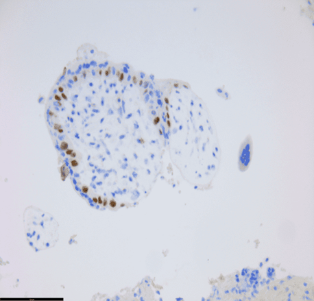
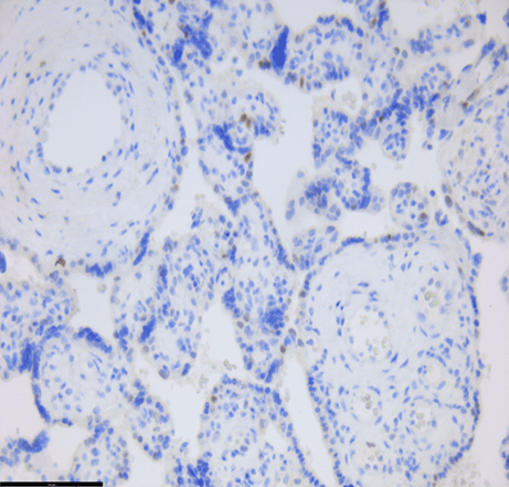
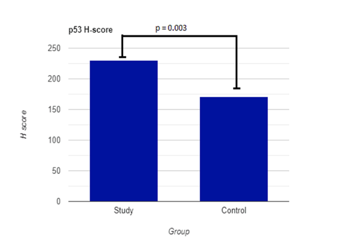
In our study, placental apoptotic changes were evaluated from the point of view of the balance between p53 expression and BCL2 expression. The analysis of pro-apoptotic versus anti-apoptotic factors in the patients included in the study shows an increased expression of p53 in the patients in the study group (Figure 1a) (230,33 ±63,80) compared to the value in the control group (Figure 1b) (171,22 ± 36,04) (Table 2). The histological and immunohistochemical analysis brings an important contribution of information both for the identification and localization of apoptosis mechanisms and for the most correct microscopic diagnosis of spontaneous abortions. In most cases of spontaneous abortion, their etiopathogenesis remains unclear or multifactorial, but in certain cases, the mechanisms involved in their production can be documented by highlighting chromogenically with the color brown for p53, and the domain of expression must be at the level of the cell nucleus. In the case of patients who had an abortion on demand, a low level of p53 expression can be observed. The low value of proapoptotic factors correlates with the survival of the product of conception and is part of the mechanisms of maternal immunogenic tolerance. When patients in the research experienced spontaneous abortions, there was a substantial increase in p53 expression.
Table 2. Bcl-2 and p53 values
|
Statistical analysis values of Bcl-2 and p53 in study group vs control group. |
|||
|
|
Study group (n=30) |
Control group (n=20) |
p |
|
p53- H score ± SD |
230,33 ± 63,80 |
171,22 ± 36,04 |
0,003 |
|
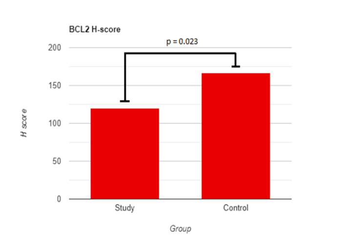
BCL2- H score ±SD |
120, ± 100 |
167 ± 92,73 |
0,023 |
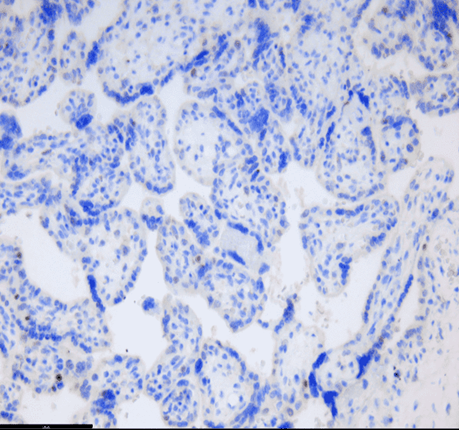
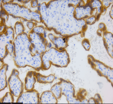
The expression analysis of the H score in the case of BCL2 reveals an increased value in the case of patients from the study group (167 ± 92,73) (Figure 1c), compared to the value obtained in the case of patients who had spontaneous abortion (120,20 ± 100) (Figure 1d).
The T-test evaluates the median of the two parameters (p53 and Bcl-2), compared with the control group. The statistical values obtained by us in the study confirm that the results are significant.
|
|
|
|
a) |
b) |
|
|
|
|
c) |
d) |
|
Figure 1. a) Higher expression of apoptotic protein (p53) in the trophoblast cells of the chorionic villi of RM cases. b) Scattered cells that are positive for p53 in sporadic abortion cases. c) Low expression of anti-apoptotic protein (Bcl-2) in the trophoblast cells of the chorionic villi of RM cases. d) High expression of Bcl-2 in sporadic abortion cases. |
|
|
|
|
Figure 2. p53 Study and control group values |
|
|
|
Figure 3. BCL2 study and control group values |
The process of programmed cell death, including apoptosis, is characterized by a sequence of complex events, which present numerous control mechanisms.
Cell proliferation, differentiation, and preservation of cell functions interfere with apoptosis. Lately, more and more pathological conditions such as ischemic lesions, autoimmune diseases, and oncological diseases have been associated with the alteration of the apoptosis process [3].
Embryogenesis is also based on the balance between cell proliferation and programmed death. Any imbalance in plus or minus can have the effect of fetal loss [10].
The current work used p53 and Bcl-2 expression to examine the placental apoptotic and anti-apoptotic pathways in instances of spontaneous abortion. When comparing the placental tissues of the study group to the control cases, there was a substantial rise in the expression of the apoptotic p53 protein and a corresponding decrease in the Bcl-2 antibodies. These findings are in line with our results, and p53's high expression is in line with other data from the literature [11, 12].
The ratio between the H score of p53 expression in the study group compared to the control group reveals an increase of over 50%. In the case of BCL2 expression, only the difference of approximately 30% in favor of the control group is noticeable. In case of decreased Bcl-2 expression, the differentiation and proliferation processes necessary for embryo-fetal evolution will be affected and the patients will have a spontaneous abortion. Through immunohistochemical methods, the inversely proportional expressions of pro- and anti-apoptotic factors (p53 vs Bcl-2) can be highlighted. The value of our study also lies in the identification of the mechanisms that led to spontaneous abortion, as it is known that there are multiple etiological factors. Diffuse, generalized cytoplasmic expression of the Bcl-2 gene is observed in the normal development of the conception product. The DAB chromogen will produce a brown color reaction by binding to the antigen-antibody complex.
Some studies have shown the reversibility of the apoptosis process under certain conditions, especially in the early stages of apoptosis induced by p53. Early repair of altered cellular DNA in the p53-induced apoptotic process can determine reversing the cell death pathway [3]. Apoptosis interferes with tumor genesis and therapies [13, 14].
The Bcl/Bax gene family is actively involved in cell escape from the apoptotic pathway. Bcl-2 expression correlates with the decrease of proapoptotic mechanisms and the augmentation of proliferative processes. In the case of the development of the product of conception, these proliferative phenomena are especially necessary during the period of organogenesis.
Our results are similar to those of other studies that demonstrate the correlation between the significantly increased level of the pro-apoptotic gene Bax in early spontaneous abortion compared to voluntarily terminated pregnancy. The Bax gene activates the expression of p53 and the induction of apoptosis via the mitochondrial pathway. Chronic endometritis is associated with infertility and spontaneous abortion. The statistical relationship between this pathology and apoptosis is not studied. Recent studies propose a screening for infertile patients [15, 16].
In the setting of miscarriage, an imbalance between pro-apoptotic and anti-apoptotic factors such as p53 and Bcl-2 may contribute to pregnancy loss. High levels of P53 can lead to increased apoptosis of trophoblast cells, which are essential for placental development and pregnancy maintenance. Conversely, low levels of Bcl-2 may result in a reduced ability to protect these cells from apoptosis. In summary, the relationship between P53 and Bcl-2 in miscarriage involves a delicate balance between cell death and cell survival. Disruption of this balance can lead to the loss of trophoblast cells and ultimately to pregnancy loss.
In the evaluation of the parameters involved in apoptotic processes (p53 and Bcl-2), we can observe an inversely proportional progression. If in the case of the study group, the value of p53 is higher compared to the control group, in the case of Bcl-2 a lower value can be observed in the spontaneous abortion compared to the medically induced abortion. All these results confirm the hypotheses studied, that the increase in apoptosis induces spontaneous abortions. The obtained results were statistically significant. The limitations of our study are the fact that it is a unicentric prospective study and the number of cases will be limited, which will lower the statistical power of conclusions.
Conclusion
Different diseases such as chronic endometritis or endometriosis can induce infertility and spontaneous abortion in close correlation with the increase in apoptotic processes. Identifying potential biomarkers corelated to the transformation of apoptotic processes from physiological processes responsible for the body's homeostasis into pathological processes associated with disease is essential for a better prognosis and for the use of personalized therapies in the future.
The diminished presence of pro-apoptotic factors is associated with the viability of the fetal product contributes to the mechanisms fostering maternal immunogenic tolerance. The examination of pro-apoptotic versus anti-apoptotic factors among the study’s participants reveal enhanced p53 expression within the study group's patients. During the assessment of parameters implicated in apoptotic processes, namely p53 and Bcl-2, a noticeable trend of inverse proportionality becomes apparent. Specifically, within the study group, there is a heightened level of p53 compared to the control group, whereas Bcl-2 levels exhibit a reduction in spontaneous abortions compared to medically induced ones. These findings corroborate the investigated hypotheses, suggesting that elevated apoptosis rates contribute to the occurrence of spontaneous abortions.
Overexpression of pro-apoptotic factors in association with the decrease in the level of anti-apoptotic factors is associated with the involution of pregnancy and the occurrence of spontaneous abortion.
Acknowledgments: None
Conflict of interest: None
Financial support: None
Ethics statement: The study was conducted in accordance with the Declaration of Helsinki, and approved by the Ethics Committee of the Resident Laboratory (protocol code 06/31 March 2020) for studies involving humans. Written informed consent has been obtained from the patient.