Pharmacophore an International Research Journal
The problem of cataract prevalence makes it necessary to develop innovative technologies for eye microsurgery. An innovative hydrodynamic scheme has been developed by a team of researchers. Its essence lies in the fact that when the vacuum level in the aspiration line reaches critical figures and remains at this level for a specified time, the system recognizes this state as occlusion and smoothly reduces the vacuum to a preset value while maintaining occlusion. The purpose of this scientific study is to study morphological changes in the cornea of rabbits' eyes after experimental modeling of a post-occlusion wave using a new hydrodynamic scheme of a phaco-emulsifier. The study was conducted on 32 eyes of 16 white laboratory rabbits, using traditional technology and an innovative hydrodynamic scheme, as well as needles of various diameters. The pieces of the cornea were carefully examined using a light microscope. The least damage to the cornea of the eye was found in the fourth group of rabbits, where an innovative hydrodynamic scheme was used in combination with a thin needle d = 0.9 mm.
Introduction
The rapid development of cataracts in modern society makes it urgent to search for innovative technological approaches that minimize surgical trauma to achieve a full rehabilitation effect in the shortest possible time [1-3]. One of the basic principles in surgical interventions on the eye is the principle of hydrodynamics of phacoemulsification, that is, the balance of inflow into the eye and outflow of fluid from the eye [4, 5]. All manipulations must take place in a stable environment with constant intraocular pressure [6, 7].
The design of phaco-emulsifiers is constantly being improved however, despite the technical and software improvements, several problems need to be solved [8-10]. One of them is the reduction of the traumatic hydrodynamic effect on the eye tissue, which can be caused by both increased fluid pressure and the collapse of the anterior chamber of the eye due to a sharp decrease in intraocular pressure [11, 12]. Sudden changes in intraocular pressure are especially dangerous since they adversely affect the state of the corneal endothelium and lens capsule, and the presence of concomitant ocular pathology (high myopia, macular degeneration, advanced glaucoma, etc.) can provoke its progression [13]. Ultimately, this leads to various intra- and post-operative complications.
Experimental evidence has proven that a breakthrough of occlusion during surgery occurs when a fragment of the lens passes through the aspiration channel of an ultrasonic or aspiration handle [14].
The authors' team has developed a new hydrodynamic scheme that changes the operating mode of vacuum automation depending on the characteristics of the aspiration flow. The essence of this method is that when the vacuum level in the aspiration line reaches critical figures and remains at this level for a specified time, the system recognizes this state as occlusion and smoothly reduces the vacuum to a preset value while maintaining occlusion. With the subsequent disappearance of occlusion, the amount of excessively aspirated liquid decreases, and, as a result, the amplitude of the pressure drop decreases.
Thus, the purpose of this scientific work is to study morphological changes in the cornea of rabbits after experimental modeling of a post-occlusion wave using a new hydrodynamic scheme for a phaco-emulsifier.
Materials and Methods
This study was performed on 32 eyes of 16 white laboratory rabbits. The animals were kept in standard, strictly identical conditions, with a standard diet. The experiments were conducted in compliance with generally accepted principles of humanity and existing international regulatory documents and instructions for working with laboratory animals [15].
Four series of experiments were performed. The animals were divided into 4 groups, respectively, they studied the degree of influence on the cornea of fluctuations in intraocular pressure during the breakthrough of occlusion. In group 1, a standard hydrodynamic scheme with a 1.1 mm needle for coaxial phacoemulsification was used, in group 2 – a new hydrodynamic scheme with a similar needle, in group 3 – a standard hydrodynamic scheme with a 0.9 mm needle for micro-coaxial phacoemulsification and in group 4 – a new hydrodynamic scheme with a 0.9 mm needle. Experimental operations were performed through incisions of 2.75 mm (groups 1 and 2) and 2.2 mm (groups 3 and 4).
The control was the intact cornea of paired rabbit eyes, samples of which were prepared for microscopic studies in parallel under identical conditions.
The procedure of surgery on laboratory animals was standard [16]. The rabbits were operated under anesthesia using Zoletil. Additionally, anesthesia was performed by injecting 1.5 ml of 2% lidocaine solution into the subtenon space, the conjunctival cavity was irrigated with 1% dicaine solution. A tunnel incision of the cornea from the temporal side was performed with a steel-calibrated peduncle with a width of 2.75 or 2.2 mm. An ultrasonic tip was inserted through a tunnel into the anterior chamber of the eye parallel to the iris and placed in the center of the pupil. At the same time, the position of the phacoagle was controlled so that it was located between the cornea and the anterior capsule, directly above the latter, and did not touch or injure the endothelium and capsule bag. To exclude the fact of accidental damage to the endothelium by crystalline masses, we deliberately refused to emulsify the lens.
The operation parameters were as follows: the height of the container with irrigation solution above eye level was 110 cm, the aspiration value was 45 ml/min, and the vacuum limit was 500 mmHg. The post–occlusion wave in each experiment was modeled as follows. With the aspiration pump running, the aspiration tube was squeezed directly near the ultrasonic handle (occlusion), after reaching the maximum value of the preset vacuum (determined by the readings on the instrument panel and stopping the pump operation), after 2 seconds the tube was unclenched (occlusion breakthrough), 10 repetitions were performed in a series. A saline solution was used as an irrigation fluid. The above scheme of the experiment most fully meets the objectives of the study and allows you to standardize its conditions as much as possible.
At the end of the operation, the animals were removed from the experiment utilizing an air embolism [17]. The eyes were enucleated 20 minutes after the death of the animal. Then the drug preparation was started: the cornea was cut out with the rim of the sclera, adjacent lens, iris, and ciliary body. The latter was necessary so as not to damage the cornea at the time of its clipping. The isolated cornea was placed on a slide table and carefully separated from the above structures using tweezers.
For microscopic examination, pieces of the cornea were fixed in paraffin wax. The fixed tissue was dehydrated in alcohols of ascending concentration, conducted through an intermediate solvent (xylene), and impregnated with paraffin, sections of 5 µm were obtained. Sections for microscopic examination were prepared using a rotary microtome LKB-III (Sweden). The prepared sections were stained with hematoxylin and eosin. The prepared histological preparation was enclosed in a Canadian balm to ensure the preservation of the object's structures, color, and transparency [18]. The sections were studied in a LEICA DM2000 light microscope (Germany) at magnification from 100 to 400.
Results and Discussion
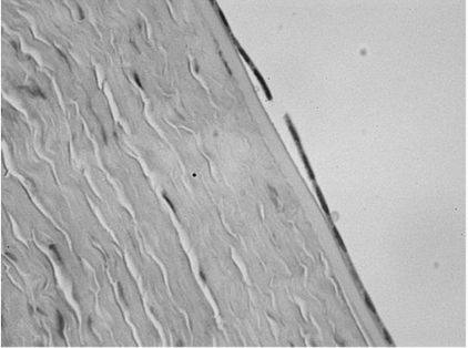
Morphology of the Cornea of Rabbits of the Control Intact Group (Norm)
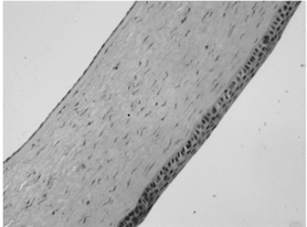
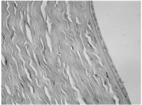
In intact rabbit eyes in the central part of the cornea, the structure of the connective tissue stroma and epithelial layers is completely preserved. Collagen fibers are arranged in dense parallel bundles, and spindle-shaped fibroblasts are visible between them. The cells of the posterior corneal epithelium lie tightly on the posterior borderline Descemet membrane without any changes (Figure 1a).
Morphology of the Cornea of Rabbits of Group 1 (Standard Hydrodynamic Scheme, Needle 1.1 mm)
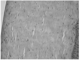
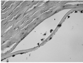
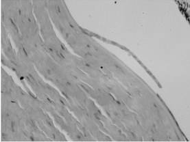
When examining the eyes of rabbits of group 1, pronounced morphological changes were found in all layers of the cornea. In the central part of the cornea, despite the integrity of the anterior multilayer epithelium, pronounced morphological changes are revealed. The connective tissue stroma of the cornea is fibrous in many areas, starting from the anterior boundary membrane and up to the posterior boundary membrane (Figure 1b). In some areas, the posterior boundary membrane of the cornea is completely separated from the connective tissue stroma (Figure 1c). At the same time, exfoliation or desquamation of many cells of the posterior single-layer epithelium from the membrane is observed.
|
|
|
|
|
a) |
b) |
c) |
|
Figure 1. The cornea of a rabbit's eye (stained with hematoxylin and eosin): a) normally, magnification x100, b) morphological changes in group 1, magnification x200, c) detachment of the posterior boundary membrane from the connective tissue stroma and desquamation of the cells of the posterior epithelium of the rabbit cornea of group 1, magnification x400 |
||
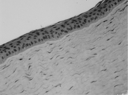
Morphology of the Cornea of Rabbits of Group 2 (New Hydrodynamic Scheme, 1.1 mm Needle)
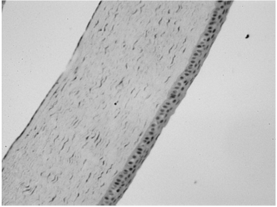
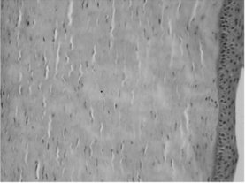
In the central part of the rabbit cornea, the structure of the anterior multilayer epithelium remains unchanged (Figure 2a). The same figure shows that in the connective tissue stroma lying directly below it, bundles of collagen fibers lie tightly and without any morphological changes. On the side of the posterior boundary membrane, weak fibrous separation of individual bundles of collagen fibers is detected in some areas, as well as swelling of the cells of the posterior epithelium in these areas or small tears in it (Figures 2b and 2c).
|
|
|
|
|
a) |
b) |
c) |
|
Figure 2. The cornea of the rabbit eye of group 2 (stained with hematoxylin and eosin): a) the structure of the central part, magnification ×100, b) the fibrous separation of individual bundles of collagen fibers and swelling of the cells of the posterior epithelium in the central part, magnification ×400, c) rupture of the layer of cells of the posterior epithelium in the central part, magnification ×400 |
||
Morphology of the Cornea of Rabbits of 3 Groups (Standard Hydrodynamic Scheme, 0.9 mm Needle)
Morphological changes are detected in the central part of the rabbit cornea, despite the integrity of the anterior multilayer epithelium. Bundles of collagen fibers of the connective tissue plate of the cornea are fibrous in many areas (Figure 3a).
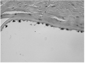
The posterior boundary membrane of the cornea in some areas peels off from the connective tissue stroma, and the cells of the posterior epithelium undergo swelling and partial or complete desquamation (Figures 3b and 3c).
|
|
|
|
|
a) |
b) |
c) |
|
Figure 3. The cornea of the rabbit eye of group 3 (stained with hematoxylin and eosin): a) morphological changes in the form of fibrous stroma in the central part, magnification ×200, b) peeling of the posterior boundary membrane from the connective tissue stroma in the central part, magnification ×400, c) desquamation of the cells of the posterior epithelium in the central part, magnification ×400 |
||
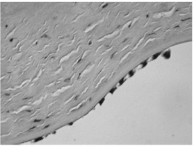
Morphology of the Cornea of Rabbits of Group 4 (New Hydrodynamic Scheme, 0.9 mm Needle)
In the central part of the rabbit cornea, the structure of the anterior multilayer epithelium is unchanged (Figure 4a). In the connective tissue stroma of the cornea, collagen fibers lie in dense parallel bundles. From the side of the posterior boundary membrane, only in some areas, weak fibrous separation of individual bundles of collagen fibers is detected, as well as opposite to these areas - swelling of cells of the posterior epithelium layer or single small tears in it (Figure 4b).
|
|
|
|
a) |
b) |
|
Figure 4. The cornea of the rabbit eye of group 4 (stained with hematoxylin and eosin): a) the structure of the anterior multilayer epithelium and stroma in the central part, magnification ×400, b) rupture of the layer of cells of the posterior epithelium in the central part, magnification ×400 |
|
Conclusion
Four series of experiments were conducted on white laboratory rabbits. The criteria for evaluating the results were changes in the anterior and posterior epithelium of the cornea, collagen fibers of the stroma, as well as damage to the posterior boundary (Descemet) membrane, as one of the most reactive structures of the cornea.
Experimental studies of the cornea of rabbits after modeling the post-occlusion wave showed that the smallest morphological changes are observed when using a micro-coaxial system with a 0.9 mm needle in combination with an innovative vacuum technology with a hydrodynamic circuit.
Acknowledgments: None
Conflict of interest: None
Financial support: None
Ethics statement: The protocol for experiments with laboratory animals complied with the requirements of the European Convention for the Protection of Vertebrate Animals used for Experimental and other Scientific Purposes.